
Virology — MCQs

On this page
What is the primary mechanism by which the influenza virus changes antigenically over time?
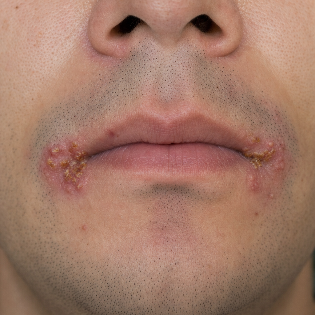
A patient presents with painful blisters around the angle of the mouth. What is the most likely pathogen responsible for this condition?
Which disease is caused by Coxsackievirus A16?
Capsid of viral structure is:
Which of the following conditions is NOT typically caused by Respiratory Syncytial Virus (RSV)?
Ebola virus belongs to?
Which is the most common influenza virus type causing disease in humans?
What is the most clinically significant common characteristic of rotavirus and Norwalk virus?
Which of the following is Hepadnavirus?
Hand foot mouth disease is caused by ?
Practice by Chapter
Virus Structure and Classification
Practice Questions
Viral Replication
Practice Questions
Pathogenesis of Viral Infections
Practice Questions
DNA Viruses: Herpesviruses
Practice Questions
DNA Viruses: Poxviruses and Adenoviruses
Practice Questions
Hepatitis Viruses
Practice Questions
RNA Viruses: Orthomyxoviruses
Practice Questions
RNA Viruses: Paramyxoviruses
Practice Questions
Enteroviruses and Rhinoviruses
Practice Questions
Arboviruses
Practice Questions
HIV and Retroviruses
Practice Questions
Oncogenic Viruses
Practice Questions
Want unlimited practice?
Get full access to all questions, explanations, and performance tracking.
Scan to download app