
Virology — MCQs

On this page
Herpangina is caused by the virus -
Cataracts and PDA in a newborn suggests in utero infection with which viral family?
Hand-foot-mouth disease is caused by:
Most commonly associated human papilloma virus with Cancer Cervix is?
Periventricular calcification seen in encephalitis is due to -
Subacute sclerosing panencephalitis is a late neurological complication associated with infection due to:
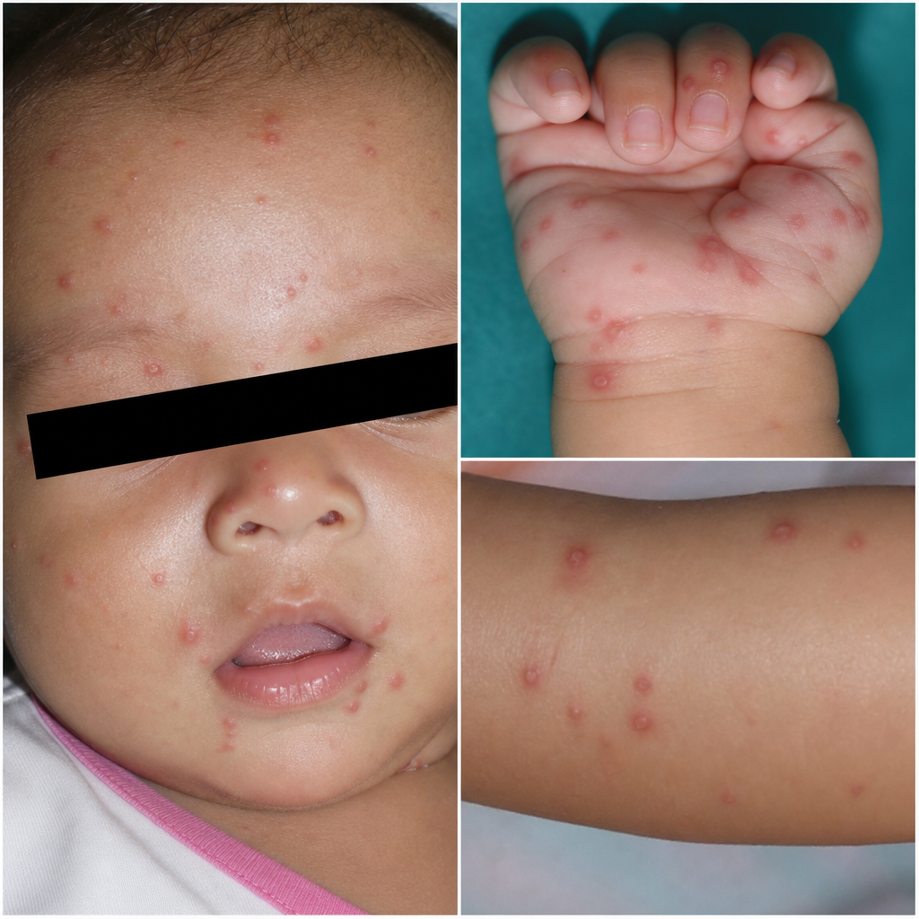
An infant presented with the following lesions on his face and limbs. Which of the following is the most likely causative organism?
The tick-borne hemorrhagic fever found in Karnataka state is:
A 5-year-old previously healthy child is taken from his day care to the pediatrician because his mother is concerned that the child has been fussy for the past few days and now has a rash on his face and torso. The mother also says the boy told her his head hurts, and she thinks he may have a low-grade fever. On examination the child is afebrile and not ill-appearing. He has a rash extending over his entire body except for the palms and soles. The infectious agent causing this disease can lead to what complication in immunocompromised hosts?
What is true about gp120?
Practice by Chapter
Virus Structure and Classification
Practice Questions
Viral Replication
Practice Questions
Pathogenesis of Viral Infections
Practice Questions
DNA Viruses: Herpesviruses
Practice Questions
DNA Viruses: Poxviruses and Adenoviruses
Practice Questions
Hepatitis Viruses
Practice Questions
RNA Viruses: Orthomyxoviruses
Practice Questions
RNA Viruses: Paramyxoviruses
Practice Questions
Enteroviruses and Rhinoviruses
Practice Questions
Arboviruses
Practice Questions
HIV and Retroviruses
Practice Questions
Oncogenic Viruses
Practice Questions
Want unlimited practice?
Get full access to all questions, explanations, and performance tracking.
Scan to download app