
Virology — MCQs

On this page
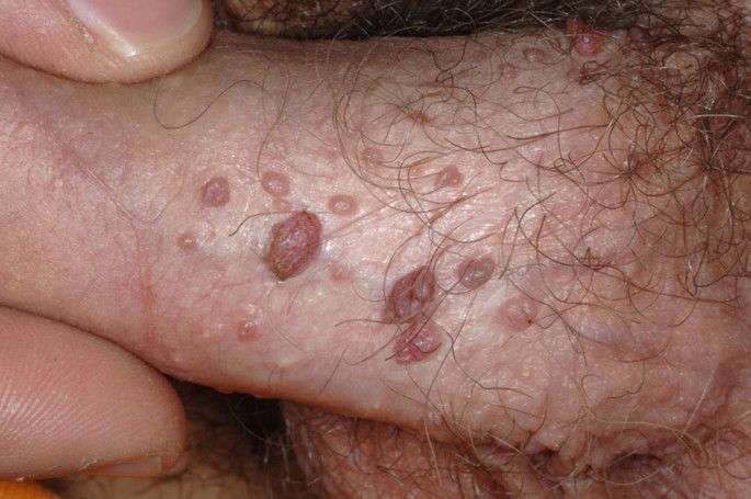
What is the causative agent for a lesion on the penis?
What change does HPV cause in the cervical epithelium?
Which of the following is NOT true about hepatitis C virus?
What is the most common cause of acute aseptic meningitis in children?
Which of the following hepatitis viruses is non-enveloped?
Human metapneumovirus is structurally similar to which of the following viruses?
What is the most important marker in diagnosing acute Hepatitis B?
Mad Cow disease (Spongiform disease) occurs due to which of the following?
Epstein-Barr virus (EBV) is associated with which of the following conditions, EXCEPT?
A 3-month-old child was admitted with pneumonia. The microscopy shows the following image. Choose the correct organism.
Practice by Chapter
Virus Structure and Classification
Practice Questions
Viral Replication
Practice Questions
Pathogenesis of Viral Infections
Practice Questions
DNA Viruses: Herpesviruses
Practice Questions
DNA Viruses: Poxviruses and Adenoviruses
Practice Questions
Hepatitis Viruses
Practice Questions
RNA Viruses: Orthomyxoviruses
Practice Questions
RNA Viruses: Paramyxoviruses
Practice Questions
Enteroviruses and Rhinoviruses
Practice Questions
Arboviruses
Practice Questions
HIV and Retroviruses
Practice Questions
Oncogenic Viruses
Practice Questions
Want unlimited practice?
Get full access to all questions, explanations, and performance tracking.
Scan to download app