
Virology — MCQs

On this page
Which of the following viruses does NOT belong to Group B arboviruses?
Herpes Zoster or Shingles represents reactivation and replication of latent infection in:
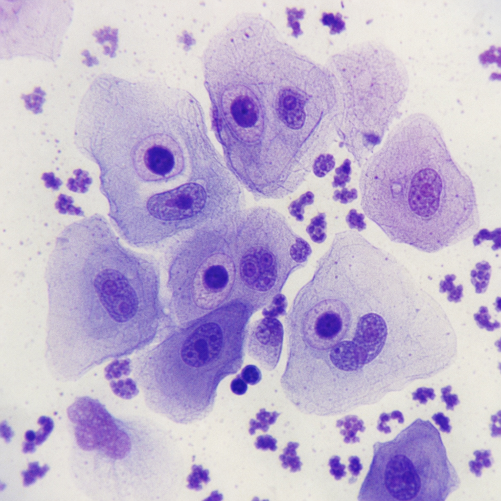
Giemsa stained smear of epithelial cells obtained from a newborn with hepatosplenomegaly is shown. What is the most likely cause of this congenital infection?
A child with agammaglobulinemia presents with respiratory tract infection and diarrhea. What is the most likely infectious agent?
A virus produces neonatal infections and infections in immunocompromised patients such as AIDS patients. The most common clinical presentation involves floaters, visual field deficits, and painless loss of vision. It can also produce encephalitis and may cause calcifying lesions in the CNS. What is this virus most likely?
A 65-year-old patient with chronic obstructive airway disease presented with headache, rhinorrhoea, myalgia, and retro-orbital and throat pain. He was managed conservatively. After 3-4 days, he presented again with cough, fever, and severe dyspnoea, requiring oxygen therapy. His condition deteriorated further, and ABG analysis showed pH 7.42, pCO2 41 mm Hg, pO2 34 mm Hg, and HCO3- 26 mmol/L. Blood cultures and sputum samples were negative for bacterial infections. Which of the following statements about the causative organism of this condition is FALSE?
Which of the following is associated with nasopharyngeal cancer?
Fatal familial insomnia is associated with which type of disease?
All are true about hepatitis viruses EXCEPT?
Elevated IgG and IgM antibody titers to parvovirus suggest a diagnosis of which one of the following?
Practice by Chapter
Virus Structure and Classification
Practice Questions
Viral Replication
Practice Questions
Pathogenesis of Viral Infections
Practice Questions
DNA Viruses: Herpesviruses
Practice Questions
DNA Viruses: Poxviruses and Adenoviruses
Practice Questions
Hepatitis Viruses
Practice Questions
RNA Viruses: Orthomyxoviruses
Practice Questions
RNA Viruses: Paramyxoviruses
Practice Questions
Enteroviruses and Rhinoviruses
Practice Questions
Arboviruses
Practice Questions
HIV and Retroviruses
Practice Questions
Oncogenic Viruses
Practice Questions
Want unlimited practice?
Get full access to all questions, explanations, and performance tracking.
Scan to download app