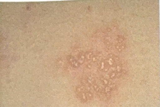
A patient presents with genital grouped vesicles, as shown in the image. What is the most likely causative organism?

Which of the following statements about herpes simplex virus (HSV) shedding is MOST accurate?
A child presented with bluish-white spots in the mouth followed by a rash. What is the genome of the most likely causative agent?
The viruses of the Filoviridae family like Ebola and Marburg resemble which of the following morphologies?
Which of the following is true about anti-CMV IgG antibodies?
A 35-year-old woman presents with painful genital ulcers that recur every few months. Current examination reveals multiple shallow ulcers on the labia minora. PCR testing is positive for HSV-2. Which of the following best explains the mechanism of viral latency in this condition?
How does HSV establish latency in the host?
Which type of HSV is most commonly associated with recurrent genital herpes?
Which cytopathic effect would confirm molluscum contagiosum virus infection?
A viral culture shows 'ground glass' nuclear appearance. Which inclusion body would confirm cytomegalovirus infection?
Explanation: ***Herpes*** - The image shows **grouped vesicles** on an erythematous base, which is the classic presentation of **genital herpes** caused by the **Herpes simplex virus (HSV)**. - These lesions are typically painful and can recur, indicating a viral etiology. *Haemophilus* - *Haemophilus ducreyi* causes **chancroid**, which presents as **painful genital ulcers** with ragged borders and often associated with inguinal lymphadenopathy. - It does not present as grouped vesicles. *Klebsiella* - *Klebsiella granulomatis* causes **donovanosis (granuloma inguinale)**, characterized by progressive, **painless ulcerative lesions** that are highly vascular and bleed easily. - This organism does not cause vesicular lesions. *Treponema* - *Treponema pallidum* causes **syphilis**, which in its primary stage presents as a **painless chancre** (a solitary ulcer) or in secondary stage as a diffuse rash. - It does not cause grouped vesicles.
Explanation: ***Asymptomatic shedding accounts for most sexual transmission*** - **Asymptomatic shedding** of HSV is very common and often goes unnoticed, leading to unsuspecting transmission to sexual partners. - Studies show that a significant proportion, often over 70%, of HSV acquisition occurs during contact with an infected partner who is **asymptomatically shedding** the virus. *HSV-1 sheds more frequently than HSV-2 in the genital tract* - While HSV-1 can cause genital herpes, **HSV-2 is the predominant cause of genital herpes** and typically sheds more frequently and for longer durations from the genital tract than HSV-1. - **Recurrent genital HSV-2 outbreaks** are also more common than recurrent genital HSV-1 outbreaks, reflecting the greater propensity for shedding. *Viral shedding occurs only during symptomatic episodes* - This statement is incorrect because **viral shedding frequently occurs in the absence of visible lesions or symptoms**, a phenomenon known as asymptomatic shedding. - Patients can shed the virus before the appearance of lesions (prodrome), during symptomatic periods, and often following resolution of symptoms or in completely **asymptomatic periods**. *Antiviral suppression completely eliminates viral shedding* - Antiviral medications like acyclovir, valacyclovir, and famciclovir can **significantly reduce the frequency and duration of HSV shedding**, both symptomatic and asymptomatic. - However, they do **not completely eliminate viral shedding**, meaning that transmission is still possible even with suppressive therapy.
Explanation: ***Enveloped virus with single-stranded RNA*** - Bluish-white spots in the mouth (**Koplik spots**) followed by a rash are pathognomonic for **measles**, which is caused by the **measles virus**. - The measles virus is a **paramyxovirus**, characterized as an **enveloped, negative-sense, single-stranded RNA virus**. *Double stranded Naked RNA* - No major human pathogen belongs to this specific genomic and structural classification. - Most **dsRNA viruses** like **rotavirus** are **naked** but cause gastroenteritis, not measles. *Naked virus with single-stranded RNA* - Viruses like **rhinovirus** (common cold) or **poliovirus** fit this description but do not cause Koplik spots or measles. - **Naked viruses** lack a lipid envelope, making them generally more resistant to environmental factors. *Double stranded Enveloped RNA* - There are no known medically significant human viruses that are both **enveloped** and contain **double-stranded RNA**. - Viral genomes are typically either DNA or RNA, and RNA viruses are usually single-stranded (positive or negative sense) or double-stranded, with or without an envelope.
Explanation: ***Filamentous*** - Viruses in the **Filoviridae family**, including **Ebola** and **Marburg**, are characterized by their distinct **long, filamentous shape**. - This morphology is a key distinguishing feature visible under electron microscopy, contributing to their namesake ("filo" meaning "thread-like"). *Brick shaped* - **Brick-shaped morphology** is characteristic of **Poxviridae**, such as the **variola virus** (smallpox). - This shape is distinctly different from the thread-like structure of filoviruses. *Bullet shaped* - **Bullet-shaped viruses** are typical of the **Rhabdoviridae family**, which includes the **rabies virus**. - This shape is consistent and easily recognizable for this family, contrasting with the much longer and flexible filaments of filoviruses. *Spherical* - **Spherical morphology** is common among many virus families, including **influenza virus** (Orthomyxoviridae) and **human immunodeficiency virus (HIV)** (Retroviridae). - While many viruses are roughly spherical, filoviruses are specifically known for their elongated, non-spherical appearance.
Explanation: ***IgG avidity assay helps in differentiating past and primary infection*** - **IgG avidity** measures the binding strength of IgG antibodies to their antigen. In a **primary infection**, IgG antibodies have low avidity. - As the immune response matures over several months, the avidity of IgG antibodies increases, indicating a **past infection**. *Denotes latent CMV infection* - While the presence of IgG antibodies indicates a past exposure and often a latent infection, it doesn't solely *denote* latency, as primary infection also involves IgG production. - **Latent CMV infection** specifically refers to the persistence of the virus in cells without active replication, which is usually confirmed by the presence of IgG antibodies but needs further contextual information like negative IgM and viral load. *Denotes chronic CMV infection with immunity to other serotypes* - CMV typically exists as one serotype, and IgG antibodies confer protection against *re-activation* of that specific virus, not immunity to "other serotypes." - **Chronic infection** usually implies ongoing active replication or persistent symptoms, which a positive IgG alone does not confirm. *Indicates acute CMV infection* - **Acute CMV infection** is primarily indicated by the presence of **IgM antibodies**, which appear early in the infection. - While IgG antibodies also rise during acute infection, their presence alone is not specific for an **acute phase** as they persist after the infection resolves.
Explanation: ***Establishment of dormancy in sensory ganglia as episomal DNA*** - HSV-2 establishes latency by maintaining its **DNA genome as a circular episome** within the **nucleus of sensory neurons** in the dorsal root ganglia (sacral ganglia for genital HSV-2). - The viral DNA remains **extrachromosomal** (not integrated) and is transcriptionally silent during latency, with only latency-associated transcripts (LATs) expressed. - Reactivation can occur due to various triggers (stress, immunosuppression, UV exposure), leading to viral replication and transport back down the axon to cause recurrent mucosal lesions. - This episomal latency mechanism is characteristic of all **herpesviruses** and allows lifelong persistence with periodic reactivation. *Integration into host genome* - This is the latency mechanism of **retroviruses** (HIV, HTLV) that use reverse transcriptase to integrate their DNA into the host chromosome. - **Herpesviruses do NOT integrate** into the host genome - they maintain episomal DNA, which is a key distinguishing feature. - HSV-2 is a DNA virus that does not require integration for persistence. *Immune evasion through antigenic drift* - **Antigenic drift** involves gradual accumulation of point mutations in surface antigens, primarily seen in **influenza virus** and allows escape from pre-existing immunity. - While HSV-2 has immune evasion mechanisms, antigenic drift is not the basis of its latency or recurrent infections. - HSV latency is established through neuronal dormancy, not through antigenic variation. *Persistent viremia* - **Persistent viremia** refers to continuous viral presence in the bloodstream, seen in chronic infections like **hepatitis B, hepatitis C, or HIV**. - HSV-2 does not cause persistent viremia; instead, it remains dormant in sensory ganglia with periodic reactivation causing localized mucosal lesions. - Between outbreaks, HSV-2 is typically not detectable in blood.
Explanation: ***Through retrograde axonal transport to dorsal root ganglia*** - HSV establishes latency by traveling via **retrograde axonal transport** from the site of infection to the sensory **dorsal root ganglia (DRG)**. - In the DRG, the virus remains dormant within the neuronal cells, expressing only latency-associated transcripts (LATs) rather than replicating. *By forming spores in epithelial cells* - HSV is a **DNA virus** and does not form spores, which are typically found in bacteria and fungi as survival structures. - While HSV infects epithelial cells initially, its latency phase is established in neurons, not by forming spores in epithelial tissues. *Through persistent viremia* - **Persistent viremia** refers to the continuous presence of viruses in the bloodstream, which is characteristic of some chronic viral infections but not how HSV establishes latency. - HSV latency involves viral DNA remaining dormant within specific neuronal cells, not actively circulating in the blood. *By integrating into host DNA* - While some viruses (like retroviruses) integrate their genetic material into the host genome, **HSV does not integrate its DNA** during latency. - HSV DNA typically remains as an **episome** (a circular, independent piece of DNA) within the nucleus of the infected neuron during latency.
Explanation: ***HSV-2*** - **HSV-2** is the primary cause of **genital herpes** and is significantly more likely to cause recurrent outbreaks than HSV-1 in the genital region. - The virus establishes **latency in sacral ganglia**, leading to frequent reactivation and subsequent genital lesions. *HSV-1* - While **HSV-1** can cause **genital herpes** (often through oral-genital contact), it is more commonly associated with **oral herpes (cold sores)**. - Genital infections caused by HSV-1 tend to recur less frequently and are generally less severe than those caused by HSV-2. *VZV (Varicella-Zoster Virus)* - **VZV** causes **chickenpox** (initial infection) and **shingles** (reactivation), not genital herpes. - It establishes latency in **dorsal root ganglia** and reactivation presents as a dermatomal rash (shingles). *CMV (Cytomegalovirus)* - **CMV** is a common virus that usually causes **asymptomatic infection** in healthy individuals, but can cause severe disease in immunocompromised patients or neonates. - It is not associated with genital lesions or recurrent genital herpes.
Explanation: ***Henderson-Paterson bodies*** - These are characteristic **large, eosinophilic intracytoplasmic inclusion bodies** seen in keratinocytes infected with the **molluscum contagiosum virus**. - Their presence confirms a diagnosis of molluscum contagiosum upon histological examination. *Cowdry type B inclusions* - These are **intracytoplasmic inclusion bodies** associated with **adenovirus** and **poliovirus** infections. - They are not characteristic of molluscum contagiosum virus, which forms distinct Henderson-Paterson bodies. *Owl's eye inclusions* - These are large, **intranuclear inclusion bodies with a clear halo** surrounding a dense central core, classically seen in cells infected with **cytomegalovirus (CMV)**. - They are distinct from the cytoplasmic inclusions of molluscum contagiosum. *Negri bodies* - These are **eosinophilic cytoplasmic inclusions** found in the cytoplasm of hippocampal pyramidal cells and Purkinje cells of the cerebellum, pathognomonic for **rabies virus infection**. - They are associated with a different viral family and are distinct from molluscum contagiosum inclusions.
Explanation: ***Owl's eye inclusions*** - **Owl's eye inclusions** are characteristic large, eosinophilic intranuclear inclusions with a clear halo, pathognomonic for **cytomegalovirus (CMV)** infection. - The "ground glass" appearance observed in a viral culture is often an early indicator, and these inclusions confirm the presence of **CMV**. *Negri bodies* - **Negri bodies** are eosinophilic **intracytoplasmic** inclusions found in the pyramidal cells of the hippocampus and Purkinje cells of the cerebellum in individuals with **rabies virus** infection. - They are unrelated to CMV and would not be present in a CMV infection. *Cowdry type A* - **Cowdry type A inclusions** are acidophilic intranuclear inclusions associated with **herpes simplex virus (HSV)** and **varicella-zoster virus (VZV)** infections. - While they are intranuclear, they are distinctly different from the 'owl's eye' appearance of CMV. *Henderson-Paterson bodies* - **Henderson-Paterson bodies** are eosinophilic intracytoplasmic inclusions found in cells infected with the **Molluscum contagiosum virus**. - These inclusions are specific to molluscum contagiosum and are not associated with CMV.
Virus Structure and Classification
Practice Questions
Viral Replication
Practice Questions
Pathogenesis of Viral Infections
Practice Questions
DNA Viruses: Herpesviruses
Practice Questions
DNA Viruses: Poxviruses and Adenoviruses
Practice Questions
Hepatitis Viruses
Practice Questions
RNA Viruses: Orthomyxoviruses
Practice Questions
RNA Viruses: Paramyxoviruses
Practice Questions
Enteroviruses and Rhinoviruses
Practice Questions
Arboviruses
Practice Questions
HIV and Retroviruses
Practice Questions
Oncogenic Viruses
Practice Questions
Get full access to all questions, explanations, and performance tracking.
Start For Free