A patient's sample is positive for infectious Hepatitis B. Choose the correct serological marker combination for this condition.
Which of the following hepatitis viruses is likely to get transmitted via fecal-oral route?
The image shown here is used for the diagnosis of
A young adult presents with facial pain and painful vesicular lesions in the mouth. Tzanck smear reveals multinucleated giant cells with intranuclear inclusions. What is the most likely causative organism?
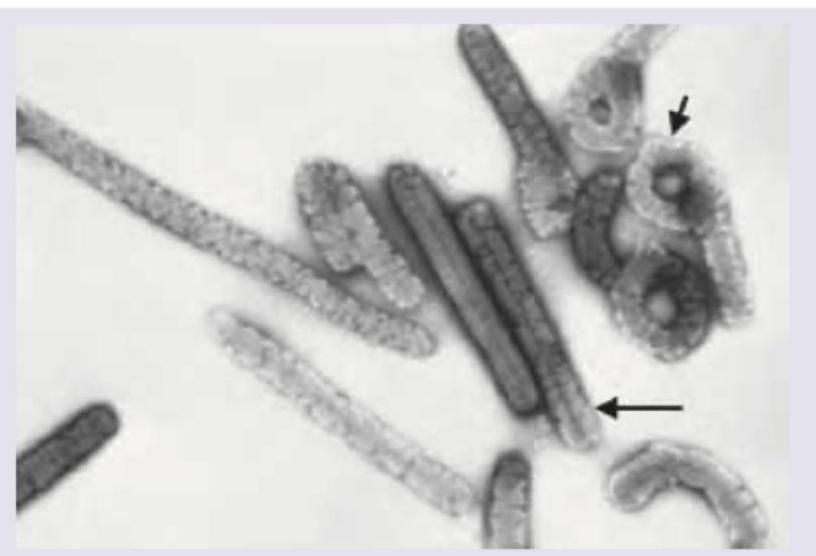
The following electron microscope image shows presence of:

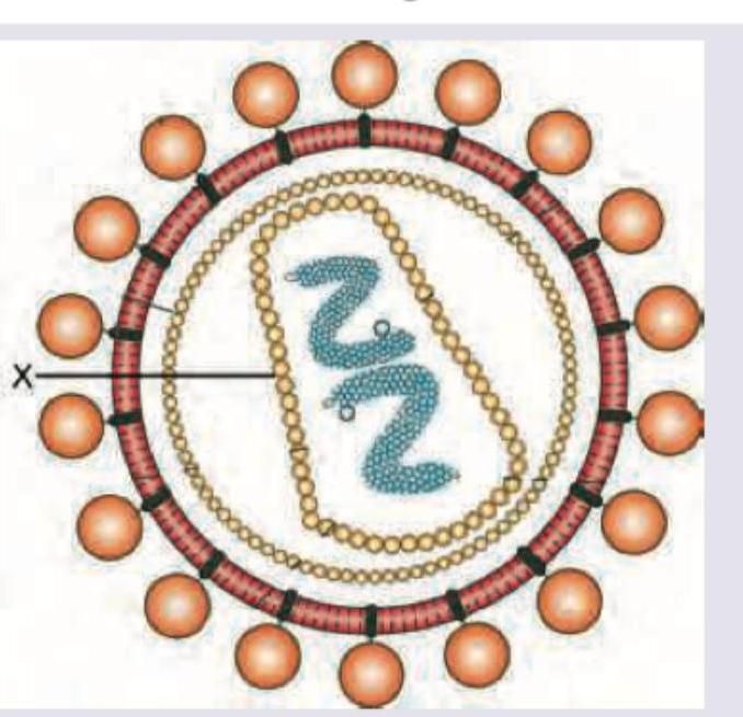
Name the antigen marked X in the figure:

Comment on the marking X in HIV virus.

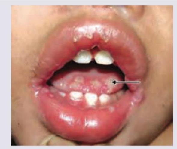
A 3-year-old child presents with fever for 3 days with excessive salivation. On examination following lesions were seen. Mother says same lesions appeared 6 months back. All are true about the condition shown except:

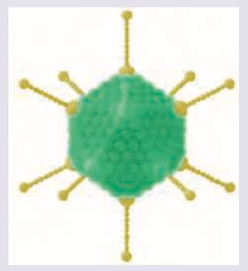
Identify the virus shown below.

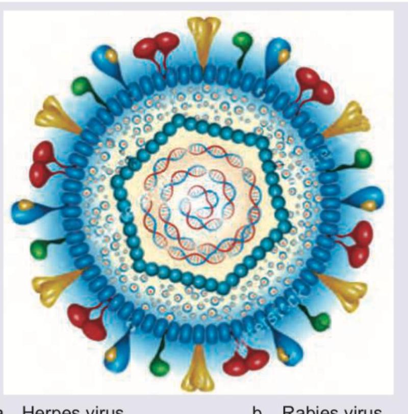
Which of the following diseases is caused by the virus shown below?

Explanation: ***Correct: HBsAg + HBeAg*** - **HBsAg (Hepatitis B surface antigen)** indicates active HBV infection (acute or chronic) - **HBeAg (Hepatitis B e antigen)** indicates active viral replication and **high infectivity** - This combination is the hallmark of **infectious/highly contagious Hepatitis B** with active virus replication - High viral load (HBV DNA levels typically >10^5 copies/mL) *Incorrect: HBcAg* - Core antigen is **not detectable in serum** - only detectable in liver tissue - Anti-HBc antibodies are detected in serum, not HBcAg itself *Incorrect: HBeAg alone* - While HBeAg indicates infectivity, **HBsAg must be present** for active infection - HBeAg without HBsAg context is incomplete for diagnosis *Incorrect: HBsAg + Anti-HBe* - This pattern indicates **low replicative phase** (inactive carrier or chronic infection with seroconversion) - Anti-HBe suggests decreased viral replication and **lower infectivity** - This is the opposite of "infectious" Hepatitis B
Explanation: ***Hepatitis A***- Hepatitis A virus (HAV) is primarily spread through the **fecal-oral route**, typically via consumption of food or water contaminated with feces.- It causes an **acute**, self-limiting infection that rarely results in chronic liver disease.*Hepatitis B*- Hepatitis B virus (HBV) is transmitted mainly through **parenteral** (exposure to infected blood), sexual contact, or perinatal (mother-to-child) routes.- Unlike HAV, HBV is a major cause of **chronic hepatitis**, cirrhosis, and hepatocellular carcinoma.*Hepatitis C*- Hepatitis C virus (HCV) transmission is predominantly **parenteral**, most commonly associated with intravenous drug use, blood transfusions (historically), and shared needles.- HCV is the leading cause of chronic viral hepatitis globally and frequently establishes persistent infection.*Hepatitis D*- Hepatitis D virus (HDV) is a defective RNA virus that requires **Hepatitis B surface antigen (HBsAg)** for its replication and assembly.- HDV is transmitted parenterally (same routes as HBV) and not via the fecal-oral route.
Explanation: ***Herpes simplex virus*** - The image is a transmission electron micrograph showing multiple enveloped virions with a characteristic dense central core and an icosahedral nucleocapsid, giving a "target" or "bull's-eye" appearance. - This morphology is a classic feature of the **Herpesviridae** family, which includes Herpes Simplex Virus (HSV), Varicella-Zoster Virus (VZV), and Cytomegalovirus (CMV). *Human immunodeficiency virus* - HIV is a **retrovirus**, and its mature virion has a distinct **cone-shaped** or **bar-shaped** core, which is morphologically different from the spherical, target-like core shown in the image. - The primary diagnostic methods for HIV are serological assays (e.g., **ELISA**) and nucleic acid tests (**PCR**), not electron microscopy. *Hepatitis B virus* - The complete infectious virion of HBV, known as the **Dane particle**, is a double-shelled spherical particle, which does not have the same internal structure as the virions in the image. - Serum from infected individuals also contains non-infectious spherical and filamentous particles of **HBsAg** which lack a nucleocapsid. *Human papillomavirus* - HPV is a **non-enveloped** DNA virus, whereas the virus in the micrograph is clearly surrounded by a lipid envelope. - It has a simple, small **icosahedral capsid** structure, lacking the complex, multi-layered appearance of a herpesvirus.
Explanation: ***Herpes simplex virus*** - The presentation of **painful vesicular lesions** in the mouth and **facial pain** strongly suggests primary (e.g., gingivostomatitis) or reactivated herpes infection. - The **Tzanck smear** showing **multinucleated giant cells** with **intranuclear inclusions** is characteristic of the Herpesviridae family, confirming the clinical suspicion of HSV. *Adenovirus* - Adenovirus typically causes **pharyngoconjunctival fever**, acute respiratory disease, or viral conjunctivitis, not localized vesicular lesions in the mouth. - It does not produce the hallmark cytopathic effect of **multinucleated giant cells** on Tzanck smear. *Cytomegalovirus* - CMV usually presents as a mild **mononucleosis-like syndrome** in healthy adults or causes severe systemic disease in the immunocompromised. - CMV histology is defined by large intranuclear **"owl's eye" inclusions**, a finding different from the multinucleated giant cells seen on Tzanck smear. *Epstein-Barr virus* - EBV causes **Infectious Mononucleosis**, characterized by fever, lymphadenopathy, and pharyngitis, not primarily painful vesicular eruptions. - Although part of the herpes family, EBV typically causes unique lymphoid pathology and **Hairy Leukoplakia**, not the cytopathic changes seen on Tzanck smear.
Explanation: ***Ebola virus*** - The electron micrograph clearly shows characteristic **filamentous, long, and pleomorphic viral particles**, some displaying coiled or branched structures, which are hallmarks of the **Filoviridae family**, to which Ebola belongs. - The presence of distinct, elongated forms, often appearing as "shepherd's crook" shapes or U-shaped structures (as indicated by the arrows), is highly indicative of the **Ebola virus morphology**. *Swine flu virus* - Swine flu, caused by influenza viruses, typically presents as **spherical or pleomorphic virions**, but not in the distinct elongated filamentous forms seen in the image. - Influenza viruses have a segmented RNA genome enclosed within a spherical capsid and an outer envelope, giving them a more rounded appearance. *Rabies virus* - Rabies virus has a classical **bullet-shaped morphology**, which is distinctly different from the long, filamentous structures observed in the image. - Its unique shape is a key identifying feature in electron microscopy. *Polio virus* - Poliovirus is a non-enveloped RNA virus with an **icosahedral (spherical) capsid structure**, much smaller and more geometrically regular than the filamentous particles shown. - It does not exhibit the elongated, flexible morphology characteristic of filoviruses.
Explanation: ***Gp120*** - The image depicts the structure of the **Human Immunodeficiency Virus (HIV)**. The antigen marked 'X' is the **outer glycoprotein** that aids in viral attachment. - **Gp120** is an exposed surface glycoprotein on HIV that binds to the **CD4 receptor** on host T-cells and other immune cells. *P24 antigen* - **P24** is a **capsid protein** found within the core of the HIV virus, not on its outer surface. - It is a crucial viral protein that encases the viral RNA and enzymes. *Gp41* - **Gp41** is a **transmembrane glycoprotein** embedded in the viral envelope, acting as an anchor for Gp120. - While associated with the viral envelope, Gp41 is mostly hidden until Gp120 binds to the host cell, facilitating **membrane fusion**. *Hemagglutinin* - **Hemagglutinin** is a **surface glycoprotein** found on the influenza virus, responsible for binding to sialic acid receptors on host cells. - It is not a component of the HIV virus.
Explanation: ***Gp120*** - The marking "X" points to the **outermost glycoprotein knob** on the HIV envelope, which is the **Gp120** subunit. - **Gp120** is crucial for attachment to host cells by binding to the **CD4 receptor** and co-receptors. *Gp41* - **Gp41** is a **transmembrane glycoprotein** that anchors Gp120 to the viral envelope and mediates membrane fusion. - It is located *underneath* Gp120, not the outermost knob. *p24 antigen* - The **p24 antigen** is a **capsid protein** that forms the conical core of the HIV virus, protecting the genetic material. - It is located *inside* the viral envelope, not on the surface. *p17 antigen* - The **p17 antigen** (also referred to as **matrix protein** or **MA**) forms a layer *just beneath* the viral envelope. - It helps maintain the viral structure and is not the outermost knob.
Explanation: ***Infectivity of type 1 virus is more temperature dependent than type 2 virus*** - This statement is **false** and is the correct answer for this EXCEPT question. - HSV-1 replicates optimally at **34-35°C** (lower temperature of oral mucosa), while HSV-2 replicates optimally at **37°C** (higher temperature of genital mucosa). - If anything, **HSV-2 is more temperature-sensitive**, requiring higher temperatures for optimal replication, making this statement incorrect. - There is no definitive evidence that HSV-1's infectivity is more temperature-dependent than HSV-2's. *Tzanck smear shows multinucleated giant cells* - A **Tzanck smear** is a cytodiagnostic test that reveals **multinucleated giant cells** and **intranuclear inclusions** in herpes simplex infections. - These findings are **characteristic** of HSV infections, supporting the diagnosis of vesicular eruptions. - This statement is **true**. *Primary target cell is mucoepithelial cells* - Herpes simplex virus (HSV) primarily infects **mucoepithelial cells** at the site of entry, leading to local replication and lesion formation. - These cells serve as the **initial site** for viral entry and proliferation during primary infection. - This statement is **true**. *Site of latency is trigeminal ganglia* - Following primary oral infection, HSV-1 travels via **sensory nerves** to establish latency in the **trigeminal ganglia**. - Reactivation from the trigeminal ganglia leads to **recurrent oral lesions**, such as the gingivostomatitis seen in this child. - This statement is **true**.
Explanation: ***Herpes virus*** - The image displays a virus with an **icosahedral capsid** (the blue beaded structure) surrounded by a viral envelope with various glycoproteins, all characteristic features of the Herpesvirus family. - The inner structure shows **double-stranded DNA** in a circular arrangement, consistent with the genomic structure of herpesviruses. *Rabies virus* - The rabies virus has a distinct **bullet-shaped morphology** and contains **single-stranded RNA**, which is clearly different from the depicted virus. - Its virions are typically 75 nm in diameter and 180 nm in length. *HIV* - HIV is a **retrovirus** with a conical-shaped capsid and contains **two copies of single-stranded RNA**, along with reverse transcriptase. - Its morphology and genetic material are distinct from the virus shown, which has a double-stranded DNA genome and an icosahedral capsid. *Influenza virus* - Influenza viruses have a roughly **spherical or filamentous shape** with a segmented **single-stranded RNA genome**. - Their surface is covered with hemagglutinin and neuraminidase spikes, distinct from the glycoproteins shown in the image.
Explanation: ***Swimming pool conjunctivitis*** - The image displays an **adenovirus**, characterized by its **icosahedral shape** and distinct **fiber proteins** projecting from the vertices. - Adenoviruses are a common cause of **pharyngoconjunctival fever**, often referred to as "swimming pool conjunctivitis" due to its spread in **inadequately chlorinated swimming pools**. - This is the **most characteristic disease** associated with adenovirus infection, particularly serotypes **3, 4, and 7**. *Neurodegenerative disorder* - Adenoviruses are **not associated** with neurodegenerative disorders. - Neurodegenerative conditions are typically linked to **prions, misfolded proteins**, or other viral agents like **JC virus** (progressive multifocal leukoencephalopathy). - Adenoviruses cause **acute infections**, not chronic neurodegeneration. *Solid organ graft infection* - While adenoviruses can cause severe infections in **immunocompromised patients**, including transplant recipients, this is not their most characteristic presentation. - In transplant patients, adenovirus may cause **colitis, hepatitis, or nephritis**, but these are **opportunistic infections** rather than the typical disease association. - The characteristic disease remains **conjunctivitis and respiratory infections** in immunocompetent hosts. *Solid organ graft rejection* - **Graft rejection** is an **immunological process** where the recipient's immune system attacks the transplanted organ, not a viral infection. - Adenovirus does not directly cause the mechanism of graft rejection. - While viral infections may complicate graft outcomes, rejection itself is **immune-mediated**, not infectious.
Virus Structure and Classification
Practice Questions
Viral Replication
Practice Questions
Pathogenesis of Viral Infections
Practice Questions
DNA Viruses: Herpesviruses
Practice Questions
DNA Viruses: Poxviruses and Adenoviruses
Practice Questions
Hepatitis Viruses
Practice Questions
RNA Viruses: Orthomyxoviruses
Practice Questions
RNA Viruses: Paramyxoviruses
Practice Questions
Enteroviruses and Rhinoviruses
Practice Questions
Arboviruses
Practice Questions
HIV and Retroviruses
Practice Questions
Oncogenic Viruses
Practice Questions
Get full access to all questions, explanations, and performance tracking.
Start For Free