All of the following statements are true regarding poliovirus, except:
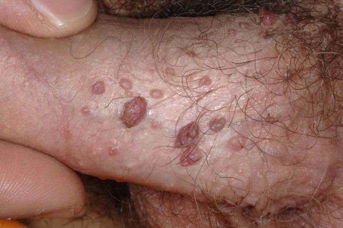
What is the causative agent for a lesion on the penis?

What change does HPV cause in the cervical epithelium?
Hepatitis E clinically resembles which other form of hepatitis?
Which of the following is associated with acute hemorrhagic conjunctivitis?
Presence of HBeAg in patients with Hepatitis B infection indicates what?
All of the following are true about Herpes group viruses except?
Which of the following viruses belongs to the Caliciviridae family?
Which influenza strain was isolated in 1989 and subsequently spread to many other countries?
A person had unprotected sex 3 weeks ago. What is the best test to rule out HIV infection at this stage?
Explanation: **Explanation:** The correct answer is **C** because Poliovirus is an Enterovirus that exists in **three distinct serotypes** (Type 1, Type 2, and Type 3). There is very little cross-immunity between these types; infection or vaccination with one type does not provide significant protection against the others. * **Type 1 (Brunhilde):** Most common cause of paralytic polio and epidemics. * **Type 2 (Lansing):** Most easily eradicated (declared eradicated globally in 2015). * **Type 3 (Leon):** Last detected in 2012 and declared eradicated in 2019. **Analysis of other options:** * **Option A:** Poliovirus is primarily transmitted via the **feco-oral route**, especially in areas with poor sanitation. It replicates in the oropharynx and intestinal Peyer’s patches. * **Option B:** In children, over **90-95% of infections are asymptomatic** (inapparent). Only about 1% of infections result in the classic paralytic disease. * **Option C:** As stated, there are three serotypes, making this statement false. * **Option D:** The **Oral Polio Vaccine (OPV/Sabin)** is a live attenuated vaccine. It induces local intestinal immunity (IgA) and is excreted in the feces, which helps immunize non-vaccinated contacts in the community, thereby creating **herd immunity**. **High-Yield Clinical Pearls for NEET-PG:** * **Specimen of choice:** Stool is the best specimen for virus isolation. * **Pathogenesis:** The virus shows tropism for the **anterior horn cells** of the spinal cord. * **Vaccine Change:** India has switched from Trivalent OPV to **Bivalent OPV** (Types 1 & 3) and introduced **Fractional IPV** to prevent Vaccine-Associated Paralytic Polio (VAPP) caused by Type 2.
Explanation: ***Human Papillomavirus (HPV)*** - HPV, particularly types 6 and 11, causes **condyloma acuminata** (genital warts), presenting as **cauliflower-like lesions** on the penis and other genital areas. - These lesions are **painless**, **flesh-colored**, and can appear as single or multiple growths, making HPV the most common cause of benign penile lesions. *Treponema pallidum* - Causes **primary syphilis** with a **painless, indurated chancre** that appears as a single, well-demarcated ulcer rather than warty growths. - The chancre has **rolled edges** and a **clean base**, distinctly different from the papillomatous appearance of condyloma acuminata. *Epstein-Barr Virus (EBV)* - Primarily causes **infectious mononucleosis** with symptoms like fever, sore throat, and **lymphadenopathy**. - Does **not typically cause penile lesions** and is associated with **nasopharyngeal carcinoma** and **Burkitt lymphoma**, not genital manifestations. *Human Herpesvirus 6 (HHV-6)* - Causes **roseola infantum** (sixth disease) in children, presenting with high fever followed by a **maculopapular rash** on the trunk. - Does **not cause penile lesions** and is not associated with sexually transmitted genital manifestations.
Explanation: **Explanation:** Human Papillomavirus (HPV), particularly high-risk types 16 and 18, is a potent oncogenic virus. The core mechanism behind its pathogenicity lies in the integration of the viral genome into the host cell DNA, leading to the overexpression of two key oncoproteins: **E6 and E7**. * **E6** binds to and degrades the **p53** tumor suppressor protein (via ubiquitin ligase). * **E7** binds to and inactivates the **Retinoblastoma (Rb)** protein, releasing the E2F transcription factor. The loss of these "guardians of the genome" prevents cell cycle arrest and DNA repair, leading to uncontrolled proliferation and the **immortalization of epithelial cells**. This transformation is the hallmark of progression from dysplasia to cervical carcinoma. **Analysis of Incorrect Options:** * **A & B (Apoptosis/Necrosis):** HPV aims to keep the host cell alive to utilize its replication machinery. E6 specifically inhibits apoptosis by degrading p53; therefore, the virus prevents rather than induces cell death. * **D (Stimulation of telomerase):** While E6 *can* indirectly activate telomerase to maintain telomere length, "Immortalization" (Option C) is the broader, more definitive pathological description of the change HPV causes in the cervical epithelium. In the context of NEET-PG, immortalization via p53/Rb inhibition is the primary recognized mechanism. **High-Yield Facts for NEET-PG:** * **Koilocytes:** Pathognomonic finding in HPV (cells with perinuclear halo and wrinkled "raisinoid" nuclei). * **Vaccination:** Quadrivalent vaccine (Gardasil) targets types 6, 11, 16, and 18. * **Screening:** Pap smear looks for cytological changes; HPV DNA testing is more sensitive for high-risk types.
Explanation: **Explanation:** Hepatitis E Virus (HEV) and Hepatitis A Virus (HAV) are clinically similar because they share the same **mode of transmission, clinical course, and lack of chronicity.** Both are "Enteric" viruses (transmitted via the **fecal-oral route**) and typically cause acute, self-limiting hepatitis. They do not lead to chronic carrier states or cirrhosis, unlike blood-borne hepatitis viruses. * **Why Hepatitis A is correct:** Both HAV and HEV present with an acute onset of fever, malaise, jaundice, and elevated transaminases. They are both non-enveloped RNA viruses (though from different families) that thrive in areas with poor sanitation and water contamination. * **Why Hepatitis B, C, and D are incorrect:** These viruses are primarily **parenteral** (blood-borne/sexually transmitted). They are characterized by their ability to cause **chronic infections**, which is not a feature of HEV. Hepatitis D specifically requires the presence of Hepatitis B (HBsAg) to replicate, a requirement not shared by HEV. **High-Yield Clinical Pearls for NEET-PG:** * **Pregnancy Risk:** While HEV is generally mild, it is notorious for causing **Fulminant Hepatic Failure in pregnant women** (especially in the 3rd trimester), with mortality rates reaching 15–25%. * **Virology:** HEV belongs to the *Hepeviridae* family. * **Zoonosis:** HEV genotype 3 is associated with the consumption of undercooked pork/deer meat. * **Chronic HEV:** Can rarely occur in immunocompromised individuals (e.g., organ transplant recipients).
Explanation: **Explanation:** **Acute Hemorrhagic Conjunctivitis (AHC)** is a highly contagious ocular infection characterized by sudden onset of painful, red eyes, subconjunctival hemorrhage, and lid edema. **Why Enterovirus is correct:** The primary causative agents of AHC are **Enterovirus 70 (EV-70)** and **Coxsackievirus A24 variant (CA24v)**. These belong to the *Picornaviridae* family. EV-70 was first identified during a major pandemic in 1969 and is specifically associated with neurological complications like polio-like paralysis (radiculomyelitis) in rare cases. **Why the other options are incorrect:** * **Rhabdovirus:** This family includes the Rabies virus, which is neurotropic and causes fatal encephalitis, not localized ocular infections. * **Calicivirus:** These viruses (e.g., Norovirus) are the leading cause of viral gastroenteritis worldwide; they do not typically cause conjunctivitis. * **Echovirus:** While Echoviruses are also Enteroviruses (Picornaviridae), they are more commonly associated with aseptic meningitis, rashes, and infantile diarrhea rather than hemorrhagic conjunctivitis. **High-Yield Clinical Pearls for NEET-PG:** * **Adenovirus (Serotypes 8, 19, 37):** These cause **Epidemic Keratoconjunctivitis (EKC)**. While EKC can show some bleeding, "Acute Hemorrhagic Conjunctivitis" as a specific clinical entity is the hallmark of EV-70 and CA24v. * **Transmission:** Fecal-oral and direct contact (hand-to-eye). * **Incubation Period:** Very short (24–48 hours), leading to explosive outbreaks in crowded settings. * **Neurological Link:** Always remember the association between EV-70 and **cranial nerve palsies or flaccid paralysis**.
Explanation: **Explanation:** The presence of **HBeAg (Hepatitis B e-antigen)** is a critical marker of **active viral replication** and high viral load. It is a soluble protein secreted by the virus into the bloodstream during the proliferative phase of the infection. **1. Why "High Infectivity" is correct:** HBeAg serves as a surrogate marker for the presence of intact virions (Dane particles) and high levels of HBV DNA. When HBeAg is detectable, the patient is highly contagious, and the risk of transmitting the virus via blood or body fluids is at its peak. Seroconversion from HBeAg to Anti-HBe usually indicates a transition to a low-replicative state. **2. Why other options are incorrect:** * **Simple carriers (Inactive carriers):** These patients are typically HBeAg negative and Anti-HBe positive, with low HBV DNA levels and normal ALT. * **Late convalescence:** This stage is characterized by the disappearance of HBsAg and the appearance of **Anti-HBs**, signifying recovery and immunity. * **Carrier status:** While a carrier can be HBeAg positive (Immunotolerant phase), the specific presence of the "e" antigen specifically denotes the *degree* of infectivity rather than just the state of being a carrier. **High-Yield Clinical Pearls for NEET-PG:** * **HBsAg:** First marker to appear; indicates current infection (acute or chronic). * **HBeAg:** Indicates active replication and **high infectivity**. * **Anti-HBc (IgM):** Best marker for the **"Window Period"** (when HBsAg and Anti-HBs are both negative). * **Anti-HBs:** Indicates **immunity** (via vaccination or recovery). * **Pre-core Mutants:** A high-yield scenario where the patient has high HBV DNA (high infectivity) but is **HBeAg negative** due to a mutation in the precore region.
Explanation: **Explanation:** The correct answer is **D**, as Burkitt’s lymphoma is a malignancy of **B cells**, not T cells. It is strongly associated with the **Epstein-Barr Virus (EBV)**, a member of the Herpesviridae family. EBV infects B cells via the CD21 receptor, leading to polyclonal B-cell proliferation and, in the presence of c-myc translocation [t(8;14)], malignant transformation. **Analysis of other options:** * **Option A (Ether sensitive):** All Herpes viruses are **enveloped** viruses. The lipid bilayer of the envelope is derived from the host nuclear membrane and is easily disrupted by organic solvents like ether or chloroform, rendering the virus non-infectious. * **Option B (May cause malignancy):** Several Herpes viruses are oncogenic. **EBV** (HHV-4) causes Burkitt’s lymphoma and Nasopharyngeal carcinoma, while **HHV-8** causes Kaposi’s sarcoma. * **Option C (HSV II involves below the diaphragm):** Traditionally, HSV-1 is associated with orofacial lesions ("above the waist/diaphragm") and HSV-2 with genital herpes ("below the waist/diaphragm"). While this distinction is blurring due to changing sexual practices, it remains a classic descriptive rule for exams. **High-Yield Clinical Pearls for NEET-PG:** * **Morphology:** Double-stranded linear DNA, icosahedral capsid, and a unique **tegument** layer between the capsid and envelope. * **Latency:** A hallmark of this family. HSV-1/2 remain latent in sensory ganglia (Trigeminal/Sacral), VZV in dorsal root ganglia, and EBV in B cells. * **Tzanck Smear:** Used for HSV and VZV to identify **multinucleated giant cells** with Cowdry Type A inclusion bodies. * **Burkitt’s Lymphoma Histology:** Characterized by a **"Starry sky" appearance** (tingible body macrophages against a sea of dark B cells).
Explanation: **Explanation:** The correct answer is **Hepatitis E virus (HEV)**. Historically, HEV was classified within the **Caliciviridae** family due to its structural similarities (non-enveloped, icosahedral, positive-sense ssRNA) to other caliciviruses like Norovirus. However, modern taxonomy has reclassified it into its own family, **Hepeviridae**. For the purpose of competitive exams like NEET-PG, HEV is still frequently associated with Caliciviridae in older textbooks and classic MCQ patterns. **Analysis of Incorrect Options:** * **Hepatitis B virus (HBV):** Belongs to the **Hepadnaviridae** family. It is the only DNA hepatitis virus (partially double-stranded circular DNA) and utilizes reverse transcriptase. * **Hepatitis C virus (HCV):** Belongs to the **Flaviviridae** family (Genus: Hepacivirus). It is an enveloped, positive-sense ssRNA virus. * **Hepatitis A virus (HAV):** Belongs to the **Picornaviridae** family (Genus: Hepatovirus). It is a non-enveloped, positive-sense ssRNA virus. **High-Yield Clinical Pearls for NEET-PG:** * **Transmission:** HEV is transmitted via the **fecal-oral route**, most commonly through contaminated water. * **Pregnancy Risk:** HEV infection carries a high mortality rate (up to 20%) in **pregnant women**, particularly during the third trimester, due to fulminant hepatic failure. * **Zoonosis:** HEV genotype 3 is associated with zoonotic transmission from pigs. * **Morphology:** HEV is described as having a "cup-shaped" indentation on electron microscopy, a characteristic feature of the Caliciviridae family (Latin *calix* = cup).
Explanation: **Explanation:** The correct answer is **H1N1**. While H1N1 is famously known for the 1918 "Spanish Flu" pandemic, it disappeared from human circulation in 1957. It re-emerged in 1977 (the "Russian Flu") and has since undergone various antigenic drifts. In **1989**, a specific variant of the **H1N1** strain was isolated and identified as a significant cause of seasonal outbreaks, subsequently spreading globally. This strain remains a major component of the trivalent and quadrivalent influenza vaccines. **Analysis of Incorrect Options:** * **H2N2 (Option A):** Responsible for the **1957 "Asian Flu"** pandemic. It circulated until 1968 and has not been seen in humans since. * **H3N2 (Option B):** Responsible for the **1968 "Hong Kong Flu"** pandemic. While it still circulates as a seasonal flu strain, it was not the specific strain highlighted for its 1989 global spread. * **H5N1 (Option D):** Known as **"Avian Influenza."** It caused a major outbreak in humans in 1997 (Hong Kong) but does not spread easily from human to human; it is primarily a zoonotic threat. **High-Yield Clinical Pearls for NEET-PG:** * **Antigenic Shift:** Major genetic changes (reassortment) leading to **pandemics** (e.g., H1N1 in 2009). * **Antigenic Drift:** Minor point mutations causing **epidemics** and necessitating annual vaccine updates. * **Influenza A** is the only type that causes pandemics due to its wide host range (birds, pigs, humans). * **Drug of Choice:** Oseltamivir (Neuraminidase inhibitor) is the standard treatment for both Influenza A and B.
Explanation: **Explanation:** The correct answer is **P24 antigen assay** because of the timing of the infection (3 weeks post-exposure). This period falls within the "window period," where the virus is replicating but the body has not yet produced detectable levels of antibodies. **1. Why P24 antigen assay is correct:** The p24 antigen is a structural protein of the HIV capsid. It appears in the blood as early as **1 to 3 weeks** after infection, coinciding with the peak of initial viremia. Since it appears before the host develops an antibody response (seroconversion), it is the preferred marker for diagnosing acute HIV infection during the early window period. **2. Why other options are incorrect:** * **ELISA (2nd/3rd Gen):** Traditional ELISA tests detect **antibodies** against HIV. Seroconversion typically takes 3 to 12 weeks. At 3 weeks, an antibody-only ELISA would likely yield a false negative. * **Western Blot:** This is a confirmatory test that detects specific antibodies to various HIV proteins (gp120, gp41, p24). Like ELISA, it relies on the host's immune response and is not sensitive enough for early acute infection. * **Lymph node biopsy:** While HIV persists in lymphoid tissue, biopsy is invasive and not a standard diagnostic tool for screening or ruling out acute infection. **Clinical Pearls for NEET-PG:** * **Window Period:** The time between infection and the point when a test can detect the virus. * **4th Generation ELISA:** The current "Gold Standard" for screening; it detects both **p24 antigen AND HIV antibodies**, significantly shortening the window period. * **Earliest Marker:** The very first marker to appear is **HIV-RNA** (detected by PCR) at approximately 10–12 days, followed by **p24 antigen** at 14–16 days. * **Best Screening Test:** 4th Gen ELISA. * **Best Confirmatory Test:** Traditionally Western Blot, though modern algorithms now favor HIV-1/HIV-2 differentiation immunoassays.
Virus Structure and Classification
Practice Questions
Viral Replication
Practice Questions
Pathogenesis of Viral Infections
Practice Questions
DNA Viruses: Herpesviruses
Practice Questions
DNA Viruses: Poxviruses and Adenoviruses
Practice Questions
Hepatitis Viruses
Practice Questions
RNA Viruses: Orthomyxoviruses
Practice Questions
RNA Viruses: Paramyxoviruses
Practice Questions
Enteroviruses and Rhinoviruses
Practice Questions
Arboviruses
Practice Questions
HIV and Retroviruses
Practice Questions
Oncogenic Viruses
Practice Questions
Get full access to all questions, explanations, and performance tracking.
Start For Free