KFD is transmitted by:
Japanese encephalitis is caused by which virus?
Vector for Zika virus disease is:
Patient: fever, joint pain, rash. Recent history of mosquito bite. Most likely diagnosis in urban area?
Which virus is most commonly recognized as a prototypical viral hemorrhagic fever in endemic regions of West Africa?
Gram stain of CSF shows gram-negative diplococci. Culture grows on Thayer-Martin medium. Most likely organism?
Subdural empyema is most commonly caused by:
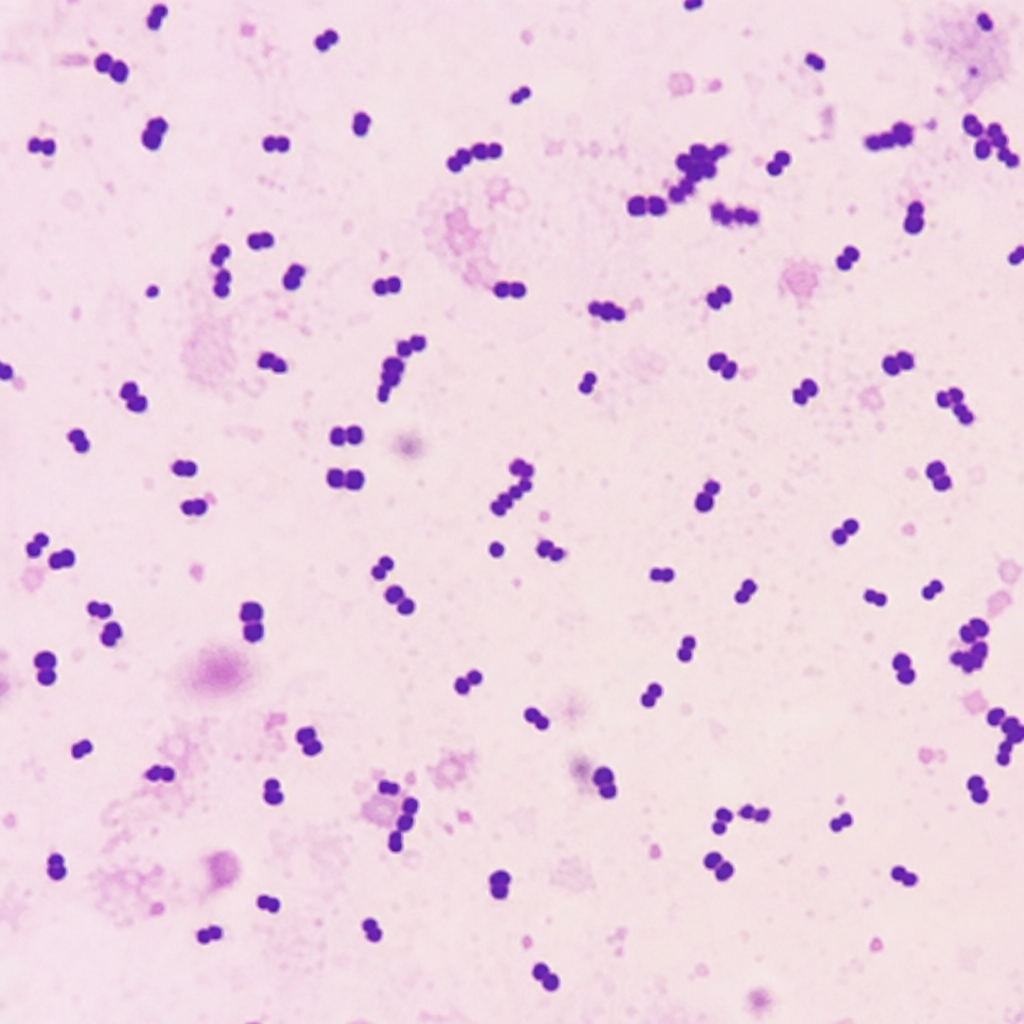
A patient presented with meningitis, and the CSF sample was subjected to gram staining and microscopy and appears as shown in the microscopic image given below. Which of the following features / tests will be characteristic of the organism?

After 5 days of birth, a baby developed poor feeding, convulsions, fever, high protein, low sugar, and low chloride levels in the cerebrospinal fluid. This is most likely due to what?
A 9-year-old child presented to OPD with complaints of high-grade fever, vomiting, and one episode of seizure. CSF examination was done and Gram staining of the culture showed lanceolate-shaped gram-positive diplococci. What is the probable causative agent?
Explanation: ***Tick*** - KFD, or **Kyasanur Forest Disease**, is a viral hemorrhagic fever endemic to India that is primarily transmitted by infected **ticks**, specifically *Haemaphysalis spinigera*. - Humans can contract the disease through a **tick bite** or contact with an **infected animal**, such as monkeys, which are reservoirs for the virus. *Sandfly* - **Sandflies** are vectors for diseases such as **leishmaniasis** and **bartonellosis**, not Kyasanur Forest Disease. - These insects transmit parasites or bacteria, whereas KFD is a **viral infection** transmitted by ticks. *Mosquito* - **Mosquitoes** are common vectors for many viral diseases, including **dengue**, **malaria**, and **chikungunya**, but they do not transmit Kyasanur Forest Disease. - KFD is exclusively associated with **tick transmission** in its endemic regions. *Mite* - **Mites** can transmit diseases like **scrub typhus** (via chiggers) or cause conditions like **scabies**, but they are not vectors for Kyasanur Forest Disease. - The primary vector for KFD is the **hard tick**.
Explanation: ***Arboviruses*** - Japanese encephalitis virus is a **flavivirus**, which belongs to the larger group of **arboviruses**. - **Arboviruses** are viruses transmitted by arthropod vectors, such as mosquitoes, which is how Japanese encephalitis spreads. *Human retrovirus* - **Retroviruses** include viruses like HIV, which are characterized by their ability to reverse-transcribe RNA into DNA. - They are not associated with encephalitis transmitted by insect vectors. *Enteroviruses* - **Enteroviruses** primarily replicate in the gastrointestinal tract and can cause a range of illnesses, including meningitis and hand-foot-and-mouth disease. - They are typically spread via the fecal-oral route, not through mosquito bites. *Cytomegalovirus* - **Cytomegalovirus (CMV)** is a herpesvirus that can cause infections in people of all ages, often asymptomatic but particularly severe in immunocompromised individuals and neonates. - CMV is transmitted through close contact, not via arthropod vectors.
Explanation: ***Aedes aegypti*** - The **Aedes aegypti mosquito** is the primary vector responsible for transmitting the Zika virus to humans. - This mosquito species is also known to transmit other arboviruses, including **dengue** and **chikungunya** viruses. *Culex* - **Culex mosquitoes** are known vectors for diseases such as **West Nile virus** and **Japanese encephalitis**. - They are generally not considered primary vectors for the Zika virus. *Phlebotomus* - **Phlebotomus (sandflies)** are vectors for parasitic diseases, most notably **leishmaniasis**. - They are not involved in the transmission of viral infections like Zika. *Anopheles stephensi* - **Anopheles mosquitoes**, particularly *Anopheles stephensi*, are the main vectors for **malaria**. - They do not play a role in the transmission of the Zika virus.
Explanation: ***Chikungunya*** - **Chikungunya** is a viral disease transmitted by mosquitoes that commonly presents with **fever**, severe **joint pain** (polyarthralgia), and a **rash**, fitting the patient's symptoms. - Its high prevalence in **urban areas** and recent history of **mosquito bites** make it a strong diagnostic consideration. *Dengue* - While Dengue also causes **fever** [1] and a **rash**, it is more typically associated with **severe muscle and bone pain** ("breakbone fever"), and **hemorrhagic manifestations** or shock, which are not mentioned. - **Joint pain** in dengue is usually less debilitating than in chikungunya. *Japanese Encephalitis* - This is a serious **neurological infection** characterized by **fever**, **headache**, seizures, and altered mental status, rather than prominent joint pain and rash. - It primarily affects the **brain** and is less likely to present with this specific symptom triad. *Malaria* - Malaria is characterized by **cyclic fevers**, chills, sweating, and fatigue, but typically **does not present with a rash** [1] or significant joint pain. - It is caused by a **parasite** transmitted by *Anopheles* mosquitoes, and its clinical picture differs from the described symptoms.
Explanation: ***Lassa fever virus*** - Lassa fever is a prototypical **viral hemorrhagic fever (VHF)** endemic to West Africa, caused by an **Arenavirus** transmitted through contact with rodent (Mastomys) excreta. - Characterized by fever, hemorrhagic manifestations in severe cases (15-20%), and multi-organ involvement with high mortality in hospitalized patients. - Represents one of the most important VHFs due to its **endemic nature** affecting millions annually in West Africa. *Yellow fever virus* - Yellow fever causes hemorrhagic fever with prominent **jaundice** and **hepatic necrosis**, transmitted by *Aedes* mosquitoes. - A **Flavivirus** causing "yellow" fever due to liver damage, but vaccine-preventable and less commonly seen in modern practice in endemic regions. *West Nile virus* - A **Flavivirus** primarily causing **neuroinvasive disease** (meningitis, encephalitis) rather than hemorrhagic fever. - Hemorrhagic manifestations are **extremely rare** and not characteristic of West Nile virus infection. *Crimean-Congo hemorrhagic fever virus* - CCHF is a severe **tick-borne VHF** (Nairovirus) with prominent hemorrhagic features and high mortality (10-40%). - Endemic to **Africa, Asia, Eastern Europe, and Middle East** but NOT West Africa, distinguishing its geographic distribution from Lassa fever.
Explanation: ***N. meningitidis*** - The presence of **gram-negative diplococci** in CSF is a hallmark feature of *Neisseria meningitidis*. - **Thayer-Martin medium** is a selective medium specifically designed for the isolation and growth of *Neisseria* species, inhibiting other bacterial and fungal contaminants. *L. monocytogenes* - *Listeria monocytogenes* is a **gram-positive rod**, which contradicts the gram stain finding of gram-negative diplococci. - It typically causes meningitis in neonates, elderly, and immunocompromised individuals, but its morphology is distinct. *S. pneumoniae* - *Streptococcus pneumoniae* is a **gram-positive coccus**, often appearing in pairs (diplococci) or short chains, but it is gram-positive, not gram-negative. - It is a common cause of bacterial meningitis but does not grow readily on Thayer-Martin medium. *H. influenzae* - *Haemophilus influenzae* is a **pleomorphic gram-negative coccobacillus**, not a diplococcus, and it requires specific growth factors (X and V factors) found in chocolate agar. - While it can cause meningitis, its characteristic morphology and growth requirements differ from the description.
Explanation: ***Streptococcus pneumoniae*** - **_Streptococcus species_**, including **_S. pneumoniae_**, aerobic and anaerobic streptococci, and S. milleri group, are the **most common causative organisms** of **subdural empyema**, accounting for **60-70% of cases**. - **Subdural empyema** most frequently arises from **paranasal sinusitis** (50-80% of cases) or **otitis media/mastoiditis**, infections typically caused by **streptococcal species**. - The polymicrobial nature of sinus and ear infections explains why **streptococci** predominate in subdural empyema. *Staphylococcus aureus* - **_Staphylococcus aureus_** is an important cause of **subdural empyema**, particularly in **post-neurosurgical cases** and following **penetrating head trauma**. - It accounts for approximately **10-20% of cases** but is **not the most common** overall pathogen. - When subdural empyema follows **surgery** or **direct inoculation**, S. aureus becomes more likely than in community-acquired cases. *H. influenzae* - **_Haemophilus influenzae_** was historically significant before widespread **Hib vaccination**. - Currently uncommon as a cause of **subdural empyema**, especially in vaccinated populations. - May still be seen in unvaccinated individuals or those with underlying immunodeficiency. *E. coli* - **_E. coli_** is a rare cause of **subdural empyema** in adults. - More relevant in **neonatal meningitis** and infections in **immunocompromised hosts**. - Gram-negative bacilli are generally uncommon in subdural empyema compared to gram-positive cocci.
Explanation: ***Catalase negative, optochin sensitive*** - The image shows **Gram-positive diplococci** (lancet-shaped, occurring in pairs), indicative of *Streptococcus pneumoniae*. - *S. pneumoniae* is known for being **catalase-negative** and **optochin-sensitive**, which are key diagnostic features that distinguish it from other α-hemolytic streptococci. - Additional characteristic: *S. pneumoniae* **ferments inulin** (bile soluble, quellung positive). *Ferments glucose and maltose but not inulin* - The phrase "does not ferment inulin" would suggest a streptococcus species OTHER than *S. pneumoniae*. - *S. pneumoniae* characteristically **DOES ferment inulin**, which is an important diagnostic feature. - This option is incorrect because it describes sugar fermentation that contradicts *S. pneumoniae* characteristics. *Oxidase and catalase negative, does not ferment inulin* - While *S. pneumoniae* is catalase-negative and oxidase-negative (oxidase test not routinely used for Gram-positive cocci), the statement "does not ferment inulin" is **incorrect** for *S. pneumoniae*. - *S. pneumoniae* **ferments inulin**, which is a key differentiating feature from other streptococci. - This combination of features does not match the organism shown in the microscopy. *Oxidase and catalase positive and does not ferment maltose or glucose* - This biochemical profile is typical for non-fermenting Gram-negative bacteria like *Pseudomonas aeruginosa*. - Such organisms would appear as **Gram-negative rods**, not the **Gram-positive diplococci** seen in the image. - This option is clearly incorrect based on the Gram stain morphology alone.
Explanation: ***Listeria monocytogenes infection*** - **Listeria meningitis** in neonates often presents with non-specific symptoms such as **poor feeding**, **convulsions**, and fever, typically in the **first week of life**. - The CSF profile of **high protein**, **low glucose**, and **low chloride** is characteristic of **bacterial meningitis**, which *Listeria* commonly causes in newborns. - *Listeria* is one of the leading causes of neonatal meningitis, particularly in early-onset sepsis (within 7 days of birth). *Tuberculosis infection* - **Tuberculous meningitis** typically has a more insidious onset and is less common in neonates unless there's significant maternal exposure. - While it can cause low glucose and high protein in CSF, the acute presentation in a 5-day-old neonate is atypical for TB. - TB meningitis has a longer incubation period and wouldn't typically present this early. *Leptospira infection* - **Leptospirosis** is rare in neonates and usually presents with symptoms like fever, headache, and muscle pain, sometimes leading to renal or hepatic involvement. - The CSF profile in leptospirosis would typically show **lymphocytic pleocytosis** with normal or slightly elevated protein, not the classic bacterial meningitis pattern. *Mycoplasma pneumoniae infection* - **Mycoplasma pneumoniae** is primarily a cause of **respiratory infections** (e.g., atypical pneumonia) in older children and adults. - It rarely causes meningitis in neonates, and neonatal infection is highly unusual. - When CNS involvement occurs, it's typically in older children with preceding respiratory symptoms.
Explanation: ***Streptococcus pneumoniae*** - The description of **lanceolate-shaped gram-positive diplococci** in CSF is characteristic of *S. pneumoniae*. - This bacterium is a common cause of **bacterial meningitis** in children and can present with high fever, vomiting, and seizures. *Haemophilus influenzae* - This is a **gram-negative coccobacillus**, which would appear as small, pleomorphic rods rather than lanceolate-shaped diplococci on Gram stain. - While it causes meningitis, its Gram stain morphology is distinct from *S. pneumoniae*. *Streptococcus agalactiae* - *S. agalactiae* (Group B Streptococcus) is a **gram-positive coccus**, but it typically appears in **chains** and is a major cause of neonatal meningitis, not usually in a 9-year-old child. - Its morphology on Gram stain would not be described as lanceolate diplococci. *Neisseria meningitidis* - *N. meningitidis* is a **gram-negative diplococcus** and would appear as kidney-bean shaped or flattened paired cocci, not gram-positive. - Though a common cause of meningitis, the Gram stain morphology described rules it out.
Get full access to all questions, explanations, and performance tracking.
Start For Free