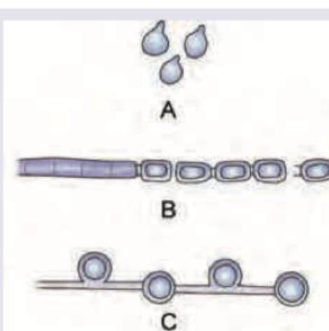
Which of the following is correct about the vegetative fungal spores?

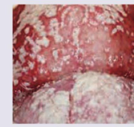
All are correct about the organism causing the following lesion except:

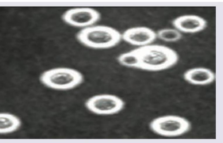
All are correct about the image shown except:

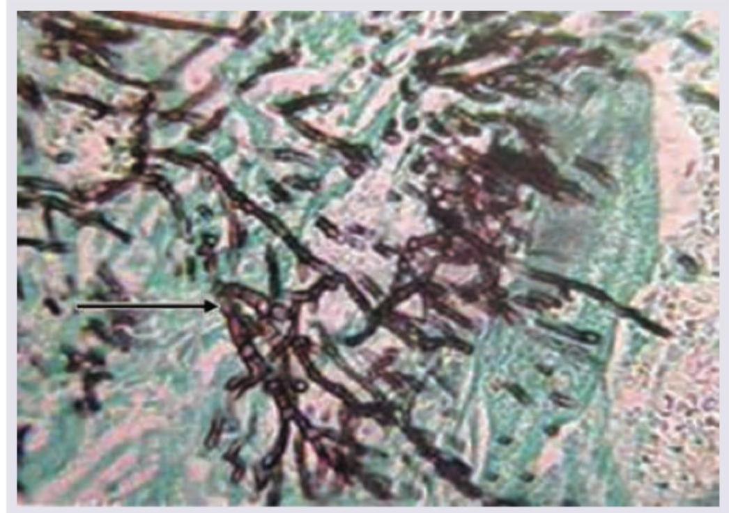
Identify the fungal organism in this slide stained with Gomori-methenamine silver stain. (AIIMS Nov 2017)

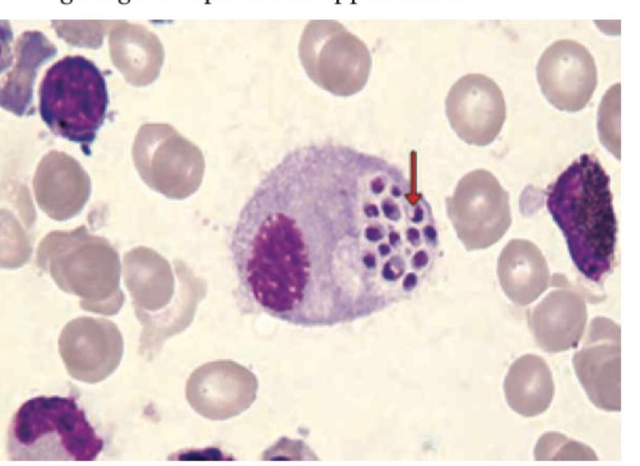
Based on the image showing intracellular yeast inside a macrophage with a peripheral crescent-shaped nucleus, which statement is true regarding Histoplasma capsulatum?

A patient in the ICU with a central venous catheter (CVC) develops an infection. Microscopy reveals ovoid budding yeast cells. What is the most likely organism?
Broad-based budding yeasts are seen in:
Desert rheumatism is caused by:
A hair perforation test was performed on a yeast isolate, and the result was positive. Which of the following organisms is associated with a positive hair perforation test?
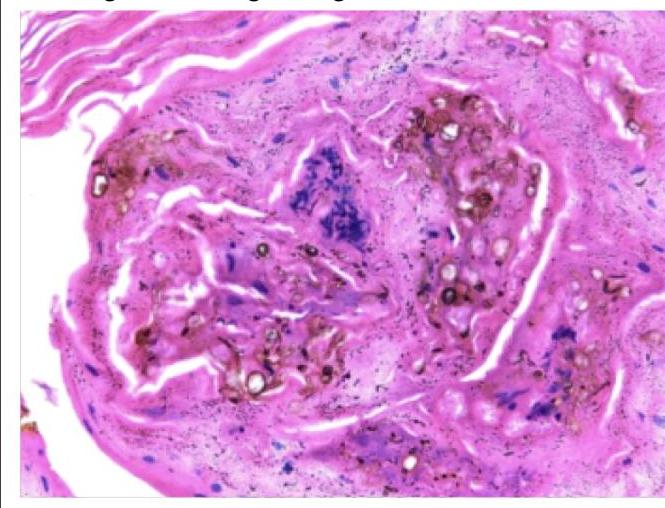
A forest worker developed skin lesions over the forearm, which initially started as macules but then became nodules. Histology of the nodule shows the following findings. Which of the following is true regarding this condition?

Explanation: **A = Blastospores, B= Arthrospores, C= Chlamydospores** - Image A depicts **blastospores**, which are asexually produced spores formed by **budding** from a parent cell, giving them a distinct tear-drop or oval shape. - Image B illustrates **arthrospores**, which are formed by the **fragmentation** of a hyphal cell into barrel-shaped segments. - Image C shows **chlamydospores**, characterized by their **thick-walled**, resistant, and usually spherical or oval structure within a hypha. *A = Arthrospores, B= Blastospores, C= Chlamydospores* - This option incorrectly identifies image A as arthrospores, which are typically barrel-shaped and result from hyphal fragmentation, not the budding pattern seen in image A. - Image B is incorrectly labeled as blastospores, but the fragmentation pattern is characteristic of arthrospores. *A = Blastospores, B= Chlamydospores, C= Arthrospores* - While image A is correctly identified as blastospores, this option misidentifies image B as chlamydospores. - Image C does not show arthrospores; the thick-walled structure is characteristic of chlamydospores, not the barrel-shaped arthrospores. *A = Chlamydospores, B= Arthrospores, C= Blastospores* - This option incorrectly identifies image A as chlamydospores, which are thick-walled resistant structures, not the budding spores visible in the image. - It also incorrectly labels image C as blastospores; the thick-walled appearance is typical of chlamydospores, not budded blastospores.
Explanation: ***Reynold Braude phenomenon*** - The **Reynolds-Braude phenomenon** is associated with **Staphylococcus aureus** and refers to the increased invasiveness of *S. aureus* in the presence of certain other bacteria. - This phenomenon is **not characteristic of *Candida albicans***, the organism responsible for the oral candidiasis (thrush) shown in the image. - **This is the correct answer** to this "except" question. *Gram positive yeast cells* - *Candida albicans* is a **Gram-positive yeast** that appears purple/blue on Gram staining. - It typically shows **budding yeast cells** and **pseudohyphae** on microscopy. - This statement is TRUE for *Candida albicans*. *Chlamydospores obtained on cornmeal agar* - **Chlamydospores** are thick-walled, large, round terminal spores that are characteristic of *Candida albicans*. - They are best demonstrated on **cornmeal agar with Tween 80** (or rice agar), which is the standard medium for their production. - The presence of chlamydospores is a key identifying feature that helps differentiate *C. albicans* from other *Candida* species. - This statement is TRUE for *Candida albicans*. *Creamy patches that on removal lead to red oozing patches* - The image shows **creamy white patches** on an inflamed oral mucosa, characteristic of **pseudomembranous candidiasis** (oral thrush). - These patches can be **scraped off**, revealing an **erythematous, bleeding** (red oozing) underlying surface. - This is a classic clinical presentation of oral candidiasis. - This statement is TRUE for *Candida albicans*.
Explanation: ***Primary site of infection is CNS*** - While *Cryptococcus neoformans* is well-known for causing **meningitis** (a CNS infection), the **primary site of infection** is typically the **lungs**, acquired through inhalation of spores. Dissemination to the CNS occurs subsequently, especially in immunocompromised individuals. *Cryptococcus neoformans* - The image, showing encapsulated yeast cells with varying sizes and budding, is characteristic of **Cryptococcus neoformans** under India ink stain, where the capsule excludes the ink, creating a halo effect. - This fungus is known for its distinctive **thick polysaccharide capsule** and its tendency to be found in environments contaminated with bird droppings. *Thick polysaccharide capsule* - The clear halo around the yeast cells in the image directly demonstrates the presence of a **thick polysaccharide capsule**, which is a key virulence factor distinguishing *Cryptococcus neoformans*. - This capsule is responsible for the organism's unique appearance in **India ink preparations** and plays a crucial role in immune evasion. *Mucicarmine stain can be used* - The **mucicarmine stain** specifically stains the **polysaccharide capsule** of *Cryptococcus neoformans* bright red, aiding in its identification in tissue samples. - This stain is a valuable diagnostic tool, particularly when dealing with tissue biopsies where the capsule might not be as distinctly visible with India ink due to cellular debris.
Explanation: **Acute angle branching, aspergillus** - The image shows **septate hyphae** with **acute angle (45-degree) branching**, which is characteristic of *Aspergillus* species. - While *Aspergillus* can be identified by conidial heads in culture, in tissue sections stained with Gomori-methenamine silver (GMS) stain, these morphological features are key. *Acute angle branching, rhizopus* - **Rhizopus** species typically exhibit **non-septate or sparsely septate hyphae** with **irregular branching**, often at wide angles. - This morphology differs from the regularly septate, acutely branching hyphae seen in the image. *Right angle branching, aspergillus* - **Aspergillus** hyphae characteristically show **acute angle (45-degree) branching**, not right-angle branching. - The hyphae are also **septate**, a feature that is clearly visible in the image. *Right angle branching, mucor* - **Mucor** species, like other Mucorales (e.g., *Rhizopus*, *Lichtheimia*), are known for their **non-septate or sparsely septate hyphae** with **wide-angle (often right-angle) branching**. - The hyphae in the image are clearly septate and branch acutely, ruling out *Mucor*.
Explanation: ***It causes disseminated disease in immunocompromised patients*** - Histoplasma capsulatum, particularly in its yeast form, can **disseminate widely** in individuals with compromised immune systems, leading to severe and life-threatening infections affecting multiple organs. - The image shows intracellular yeast within a macrophage, which is characteristic of *Histoplasma* infection and its ability to survive within phagocytic cells during dissemination. - **Classic presentation:** fever, weight loss, hepatosplenomegaly, pancytopenia in AIDS patients or other immunocompromised states. *It cannot be grown in SDA* - This statement is incorrect; *Histoplasma capsulatum* **can be grown on Sabouraud Dextrose Agar (SDA)**, although it requires longer incubation times (2-4 weeks) at 25°C. - The fungus grows as a **mold form** at room temperature with characteristic **tuberculate macroconidia**. *It is a monomorphic fungus that exists only as yeast* - This statement is **incorrect**. *Histoplasma capsulatum* is a **dimorphic fungus**, not monomorphic. - It exists as a **mold at 25°C** (in soil enriched with bird/bat droppings) and as a **yeast at 37°C** (in host tissues). - This thermal dimorphism is a defining characteristic of the organism. *Its yeast form shows broad-based budding* - This statement is incorrect. The yeast form of *Histoplasma capsulatum* is **small (2-4 μm), oval-shaped** and reproduces by **narrow-based budding**. - **Broad-based budding** is characteristic of *Blastomyces dermatitidis*, which produces larger yeasts (8-15 μm).
Explanation: ***Candida*** - **Gram-positive ovoid budding organisms** are characteristic findings for yeast, with **Candida** species being the most common cause of CVC-related fungal infections in ICU patients. - Patients with CVCs are at high risk for candidemia due to compromised skin barriers and often receiving broad-spectrum antibiotics, which can disrupt the normal flora. *Staphylococcus epidermidis* - This is a **Gram-positive coccus** that grows in clusters and is a common cause of CVC-related **bacterial infections**, developing **biofilms** on catheters. - It does not present as an ovoid budding organism on microscopy. *Escherichia coli* - This is a **Gram-negative rod**, typically associated with **urinary tract infections** and sepsis from an abdominal source. - It would not appear as a Gram-positive ovoid budding organism and is not a common cause of primary CVC-related bloodstream infections unless there's an associated abdominal source. *Staphylococcus aureus* - This is a **Gram-positive coccus** that grows in grape-like clusters and can cause severe CVC-related bloodstream infections, often leading to **endocarditis** or widespread dissemination. - Like *S. epidermidis*, it is a bacterium and does not exhibit ovoid budding.
Explanation: ***Blastomycosis*** - This fungal infection is classically characterized by **broad-based budding yeasts** seen on microscopic examination. - The yeast cells are typically large and have a characteristic wide connection between the mother and daughter cells during budding. *Histoplasmosis* - Characterized by **small, intracellular yeasts** often seen within macrophages. - These yeasts do **not exhibit broad-based budding**. *Candidiasis* - Primarily presents as **pseudohyphae** (elongated yeast cells resembling hyphae) and budding yeasts (blastoconidia) with **narrow bases**. - **True hyphae** may also be present depending on the species and growth conditions. *Coccidioidomycosis* - In tissue, it is characterized by **spherules** containing **endospores**, not budding yeasts. - The mycelial form is found in culture or environmental samples.
Explanation: ***Coccidioides immitis*** - This fungus is the causative agent of **coccidioidomycosis**, also known as **Valley fever** or **desert rheumatism**, due to its prevalence in arid regions. - Infection most commonly occurs through inhalation of **arthroconidia** from disturbed soil in endemic areas. *Paracoccidioides brasiliensis* - This fungus causes **paracoccidioidomycosis** (South American blastomycosis), which is endemic to Latin America. - It typically manifests as chronic granulomatous disease affecting the lungs, skin, mucous membranes, lymph nodes, and internal organs. *Candida albicans* - This is a common opportunistic yeast responsible for various infections, from **superficial mucocutaneous candidiasis** (e.g., thrush, vaginal yeast infections) to severe invasive candidemia. - It is not associated with "desert rheumatism." *Cryptococcus neoformans* - This encapsulated yeast is a major cause of **cryptococcosis**, particularly in immunocompromised individuals. - It commonly causes **meningoencephalitis** and pulmonary disease, and is associated with bird droppings, but not "desert rheumatism."
Explanation: ***Candida albicans*** - The **hair perforation test** is a classic diagnostic test specifically used to identify *Candida albicans*, which characteristically produces **conical projections** when incubated with sterilized blonde hair in water at room temperature. - This test has historically been used as a simple, rapid method for **presumptive identification** of *C. albicans* from other Candida species, indicating the organism's ability to produce **keratinolytic enzymes**. *Candida tropicalis* - *Candida tropicalis* gives a **negative hair perforation test** and does not produce the characteristic hair perforations seen with *C. albicans*. - While it can form **pseudohyphae** and true hyphae, it can be differentiated from *C. albicans* using this test among others. *Candida glabrata* - *Candida glabrata* does **NOT** produce a positive hair perforation test and typically remains in **yeast form**. - It is distinguished from other Candida species by its **smaller cell size** and inability to form germ tubes or hair perforations. *Candida parapsilosis* - *Candida parapsilosis* also gives a **negative hair perforation test** and does not produce hair perforations. - This species can form **pseudohyphae** but the hair perforation test helps distinguish it from *C. albicans*.
Explanation: The image displays **chromoblastomycosis**, a fungal infection characterized by **medlar bodies** or **sclerotic bodies**. These are thick-walled, septate, dematiaceous (pigmented) fungal cells that resemble copper pennies. The patient's history of being a forest worker with skin lesions progressing from macules to nodules is consistent with this diagnosis as it's often associated with **traumatic inoculation** from contaminated plant material. ***It is a dematiaceous fungus*** - The image shows **"copper pennies"** or **sclerotic bodies**, which are characteristic of dematiaceous (pigmented) fungi causing chromoblastomycosis. - These fungi contain **melanin** in their cell walls, which contributes to their characteristic dark appearance. - Common causative agents include *Fonsecaea pedrosoi*, *Phialophora verrucosa*, and *Cladophialophora carrionii*. *Angioinvasion is common especially in people with hemolytic anemia* - **Angioinvasion** is not a feature of chromoblastomycosis, which typically remains confined to the **skin and subcutaneous tissue**. - Angioinvasion is characteristic of **mucormycosis** and **aspergillosis**, particularly in immunocompromised patients, not chromoblastomycosis. *These bodies are formed by engulfment of the dead fungi by the macrophages* - The **sclerotic bodies** are **living fungal cells** in their tissue-specific form, not dead fungi engulfed by macrophages. - They are a distinct morphological form of the fungus, adapting to growth within the host tissue, and are **actively pathogenic**. - These thick-walled structures allow the fungus to persist in tissue and resist host defenses. *Infection commonly spreads to involve tendon, muscle and bone* - Chromoblastomycosis causes **chronic, localized infections** primarily of the **skin and subcutaneous tissue**. - While local tissue destruction can occur, **deep invasion** into tendons, muscles, or bones is **rare** and occurs only in severe, long-standing cases. - The infection typically remains confined to cutaneous and subcutaneous layers without dissemination.
Classification of Fungi
Practice Questions
Superficial Mycoses
Practice Questions
Dermatophytes
Practice Questions
Subcutaneous Mycoses
Practice Questions
Candidiasis
Practice Questions
Aspergillosis
Practice Questions
Cryptococcosis
Practice Questions
Zygomycosis
Practice Questions
Endemic Mycoses
Practice Questions
Opportunistic Fungal Infections
Practice Questions
Antifungal Agents
Practice Questions
Laboratory Diagnosis of Fungal Infections
Practice Questions
Get full access to all questions, explanations, and performance tracking.
Start For Free