
Mycology — MCQs

On this page
Pneumocystis jirovecii causes infection primarily in which species?
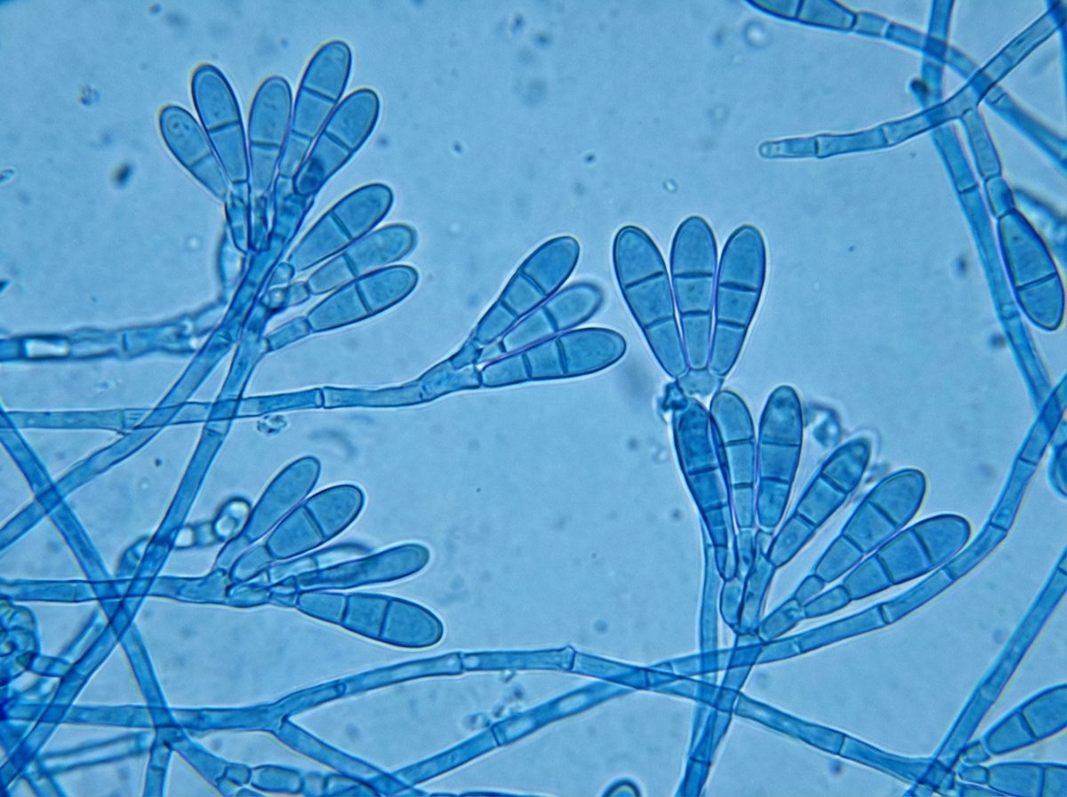
An 18-year-old white male high-school student presents with a diffuse, painful rash on his thighs extending to his navel. He reports using topical hydrocortisone from a teammate for a previous groin rash. A KOH scraping of the current lesion shows hyaline hyphae. A schematic of the microscopic observation of the culture is provided. What is the most likely etiology?
Which of the following is NOT an 'all except' characteristic of acute pseudomembranous candidiasis?
Black coloured hard nodules in hair are produced by which organism?
A 28-year-old HIV-positive male complains of pain on swallowing. Physical examination is remarkable for white plaque-like material on his tongue and buccal mucosa, which is scraped and sent to the laboratory. Based on these findings and the laboratory results, the man is diagnosed with acquired immunodeficiency syndrome (AIDS). With which of the following agents is the man most likely infected?
Which fungus cannot be grown on artificial media?
Which type of candidiasis is associated with leukoplakia?
A patient self-diagnosed with athlete's foot (tinea pedis) used an over-the-counter product. While the condition improved, it did not fully resolve. A dermatologist was consulted, and a skin scraping was sent for fungal culture. The culture yielded a slow-growing colony that produced a few small microconidia. This finding is consistent with the isolation of a dermatophyte from which genera?
Which of the following is not a fungal infection?
All statements are true about mycetoma except:
Practice by Chapter
Classification of Fungi
Practice Questions
Superficial Mycoses
Practice Questions
Dermatophytes
Practice Questions
Subcutaneous Mycoses
Practice Questions
Candidiasis
Practice Questions
Aspergillosis
Practice Questions
Cryptococcosis
Practice Questions
Zygomycosis
Practice Questions
Endemic Mycoses
Practice Questions
Opportunistic Fungal Infections
Practice Questions
Antifungal Agents
Practice Questions
Laboratory Diagnosis of Fungal Infections
Practice Questions
Want unlimited practice?
Get full access to all questions, explanations, and performance tracking.
Scan to download app