
Mycology — MCQs

On this page
Sulphur granules are composed of?
Homosexual men with AIDS are most likely to exhibit which of the following conditions?
A female presented with thick white discharge and pruritus. What is the etiological agent?
Which of the following organisms cannot be cultured?
A 38-year-old AIDS patient presents with a one-week history of fever and increasing headache. He also reports photophobia, nausea, and weakness. His past medical history is significant for Pneumocystis pneumonia, and his CD4 count is 89. Current medications include trimethoprim/sulfamethoxazole and indinavir. Cerebrospinal fluid (CSF) analysis reveals 4 WBCs, and budding encapsulated yeast forms grow on Sabouraud's agar. What is the accurate description of the morphology of the infectious form of the organism responsible for this patient's illness?
Which of the following is NOT a cause of Madura foot?
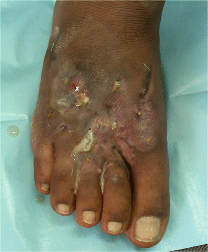
A 40-year-old man presented with swelling over his foot with multiple sinus discharge. What is the likely diagnosis?
Cigar-shaped yeast cells are seen with which of the following organisms?
A man developed a pustule after being pricked by a thorn in the garden. Laboratory examination of the tissue specimen shows cigar-shaped budding yeasts. What is the most probable causative agent?
Penicillium marneffei is commonly associated with which of the following conditions?
Practice by Chapter
Classification of Fungi
Practice Questions
Superficial Mycoses
Practice Questions
Dermatophytes
Practice Questions
Subcutaneous Mycoses
Practice Questions
Candidiasis
Practice Questions
Aspergillosis
Practice Questions
Cryptococcosis
Practice Questions
Zygomycosis
Practice Questions
Endemic Mycoses
Practice Questions
Opportunistic Fungal Infections
Practice Questions
Antifungal Agents
Practice Questions
Laboratory Diagnosis of Fungal Infections
Practice Questions
Want unlimited practice?
Get full access to all questions, explanations, and performance tracking.
Scan to download app