A 55-year-old woman presents with persistent cough, fever, and hemoptysis. Sputum shows branching septate hyphae. What is the likely pathogen?
A patient presented with some unknown fungal infection. Microscopic examination revealed brown coloured spherical fungi with septate hyphae. Possible condition:
Broad-based budding yeasts are seen in:
What type of spore is produced by Ascomycota during sexual reproduction?
Aseptate hyphae is not seen in which of the following fungi?
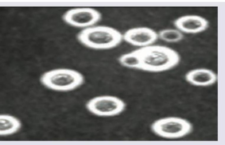
All are correct about the image shown except:

Beta 1,3 Glucan test is positive in all except?
Which statement is false regarding Cryptococcus neoformans?
Which of the following is a non-culturable fungus?
Which of the following is a dimorphic fungus?
Explanation: ***Aspergillus fumigatus*** - The presence of **branching septate hyphae** in sputum, along with symptoms of **persistent cough, fever, and hemoptysis**, is highly characteristic of an *Aspergillus* infection, particularly in immunocompromised patients or those with pre-existing lung conditions. - This fungus often colonizes the respiratory tract and can cause various diseases, including **allergic bronchopulmonary aspergillosis (ABPA)**, **aspergilloma** (fungus ball), or **invasive aspergillosis**. - The hyphae branch at **acute angles (45°)** and are **septate**, which is the key distinguishing feature. *Candida albicans* - While *Candida albicans* is a common fungal pathogen, it typically presents as **yeast** or **pseudohyphae** on microscopy, not branching septate hyphae. - It usually causes **mucocutaneous infections** like thrush or candidemia, with pulmonary involvement being less common and usually presenting differently from the described symptoms. *Histoplasma capsulatum* - *Histoplasma capsulatum* is a **dimorphic fungus** that appears as **small intracellular yeast forms** within macrophages in tissue or sputum, not branching septate hyphae. - It is endemic to certain regions (e.g., Ohio and Mississippi River valleys) and typically causes **pulmonary histoplasmosis**, which can mimic tuberculosis, but microscopic findings differ significantly. *Mucor species* - **Mucor species** are characterized by **broad, ribbon-like, aseptate (non-septate) hyphae** with irregular branching at right angles, which is distinct from the branching septate hyphae described. - These fungi typically cause **mucormycosis** (zygomycosis), an aggressive infection often seen in immunocompromised individuals, especially diabetics with ketoacidosis, and can involve the rhinocerebral region, lungs, or skin.
Explanation: ***Chromoblastomycosis*** - This infection is characterized by the presence of **sclerotic bodies** (also known as fumagoid bodies or Medlar bodies), which are **brown-pigmented**, **spherical**, copper-colored cells or muriform cells observed in tissue. - The causative fungi, often dematiaceous molds, exhibit **septate hyphae** in culture. These molds are pigmented due to melanin in their cell walls. *Histoplasmosis* - Caused by *Histoplasma capsulatum*, which appears as **small, oval, intracellular budding yeasts** within macrophages in tissue samples. - It does not typically form sclerotic bodies or prominent septate hyphae in infected tissue. *Candida albicans* - This yeast typically presents as **ovoid budding yeast cells**, pseudohyphae, and true hyphae in tissues. - It is an **achlorophyllous** fungus, meaning it lacks pigmentation and would not appear as "brown coloured" under microscopic examination. *Coccidioidomycosis* - Caused by *Coccidioides immitis* or *C. posadasii*, which are characterized by the formation of **spherules** containing endospores in tissue. - It does not form "brown coloured spherical fungi with septate hyphae" as described; spherules are large, non-pigmented, and contain smaller endospores.
Explanation: ***Blastomycosis*** - This fungal infection is classically characterized by **broad-based budding yeasts** seen on microscopic examination. - The yeast cells are typically large and have a characteristic wide connection between the mother and daughter cells during budding. *Histoplasmosis* - Characterized by **small, intracellular yeasts** often seen within macrophages. - These yeasts do **not exhibit broad-based budding**. *Candidiasis* - Primarily presents as **pseudohyphae** (elongated yeast cells resembling hyphae) and budding yeasts (blastoconidia) with **narrow bases**. - **True hyphae** may also be present depending on the species and growth conditions. *Coccidioidomycosis* - In tissue, it is characterized by **spherules** containing **endospores**, not budding yeasts. - The mycelial form is found in culture or environmental samples.
Explanation: ***Ascospores*** - **Ascospores** are the sexual spores produced by fungi belonging to the phylum **Ascomycota** during their sexual reproductive cycle. - These spores are formed inside a sac-like structure called an **ascus** after **karyogamy (nuclear fusion)** and **meiosis**. - Each ascus typically contains **4-8 ascospores** arranged in a characteristic pattern. - Examples of Ascomycota include *Aspergillus*, *Penicillium*, *Candida*, and yeasts like *Saccharomyces*. *Asexual spores* - **Asexual spores** are produced during **asexual reproduction** without the fusion of gametes or meiosis. - Examples include **conidia** and **sporangiospores**, which allow for rapid proliferation and dispersal. *Conidia* - **Conidia** are a specific type of **asexual spore**, not sexual spores. - They are formed exogenously on specialized structures called **conidiophores**. - While Ascomycota can produce conidia asexually, the question asks specifically about sexual reproduction. *None of the options* - This option is incorrect because **ascospores** are indeed the sexual spores of Ascomycota.
Explanation: ***Correct: Aspergillus*** - *Aspergillus* species are characterized by **septate hyphae** and **acute angle branching**, distinguishing them from zygomycetes. - They cause diseases such as **allergic bronchopulmonary aspergillosis (ABPA)**, aspergilloma, and invasive aspergillosis. - Since Aspergillus has septate hyphae, **aseptate hyphae are NOT seen** in Aspergillus. *Incorrect: Rhizopus* - *Rhizopus* is a zygomycete with **broad, ribbon-like, aseptate hyphae** and **right-angle/irregular branching**. - Common cause of **mucormycosis (zygomycosis)**, especially in immunocompromised individuals. *Incorrect: Mucor* - *Mucor* is another zygomycete with **aseptate hyphae** and **wide-angle branching**. - Causes mucormycosis, leading to severe infections primarily in patients with **diabetic ketoacidosis** or **hematologic malignancies**. *Incorrect: Penicillium* - *Penicillium* species possess **septate hyphae** with characteristic **brush-like (penicillus-shaped)** fruiting bodies. - While some produce antibiotics, certain species like *Talaromyces marneffei* can cause systemic infections in immunocompromised patients.
Explanation: ***Primary site of infection is CNS*** - While *Cryptococcus neoformans* is well-known for causing **meningitis** (a CNS infection), the **primary site of infection** is typically the **lungs**, acquired through inhalation of spores. Dissemination to the CNS occurs subsequently, especially in immunocompromised individuals. *Cryptococcus neoformans* - The image, showing encapsulated yeast cells with varying sizes and budding, is characteristic of **Cryptococcus neoformans** under India ink stain, where the capsule excludes the ink, creating a halo effect. - This fungus is known for its distinctive **thick polysaccharide capsule** and its tendency to be found in environments contaminated with bird droppings. *Thick polysaccharide capsule* - The clear halo around the yeast cells in the image directly demonstrates the presence of a **thick polysaccharide capsule**, which is a key virulence factor distinguishing *Cryptococcus neoformans*. - This capsule is responsible for the organism's unique appearance in **India ink preparations** and plays a crucial role in immune evasion. *Mucicarmine stain can be used* - The **mucicarmine stain** specifically stains the **polysaccharide capsule** of *Cryptococcus neoformans* bright red, aiding in its identification in tissue samples. - This stain is a valuable diagnostic tool, particularly when dealing with tissue biopsies where the capsule might not be as distinctly visible with India ink due to cellular debris.
Explanation: ***Mucormycosis*** - Fungi causing mucormycosis belong to the order **Mucorales**, which structurally lack **beta-D-glucan** in their cell walls. - Due to the absence of beta-D-glucan, the **beta-1,3-D-glucan assay** will yield a negative result in cases of mucormycosis. *Pneumocystis jirovecii* - This fungus contains significant amounts of **beta-D-glucan** in its cell wall, making the test usually positive during active infection. - A positive **beta-D-glucan test** can be a useful diagnostic marker for **Pneumocystis pneumonia (PCP)**, especially in immunocompromised patients. *Candida* - **Candida species** possess a cell wall rich in **beta-D-glucan**, leading to a positive test result during active infection. - The **beta-D-glucan assay** is a valuable adjunctive test for diagnosing invasive candidiasis. *Aspergillus* - The cell wall of **Aspergillus** contains **beta-D-glucan**, causing the test to be positive in cases of invasive aspergillosis. - A positive **beta-D-glucan test** can aid in the early diagnosis and management of invasive aspergillosis, particularly in high-risk patients.
Explanation: **Explanation:** *Cryptococcus neoformans* is an encapsulated yeast primarily associated with pigeon droppings and is a significant opportunistic pathogen in immunocompromised patients (e.g., HIV/AIDS). **1. Why "Urease negative" is the False Statement:** *Cryptococcus neoformans* is characteristically **Urease positive**. The production of the urease enzyme is a key biochemical marker used in the laboratory to differentiate it from other yeasts like *Candida albicans* (which is urease negative). It hydrolyzes urea to produce ammonia, raising the pH and changing the indicator color. **2. Analysis of Other Options:** * **Option A (Grows at 5°C and 37°C):** This is true. Unlike many other pathogenic fungi, *C. neoformans* can grow at 37°C (essential for human pathogenicity) and also at lower temperatures like 4°C–5°C. * **Option B (Has 4 serotypes):** This is true. Based on capsular polysaccharide antigens, it is classified into four serotypes: **A, B, C, and D**. (Note: Serotype A is *C. neoformans var. grubii*, while B and C are now often classified as *C. gattii*). * **Option D (Causes superficial skin infection):** This is true. While meningitis is the most common presentation, primary or secondary cutaneous cryptococcosis can occur, presenting as papules, pustules, or ulcerations. **High-Yield Clinical Pearls for NEET-PG:** * **Virulence Factor:** The **Polysaccharide capsule** (Glucuronoxylomannan) is the most important; it inhibits phagocytosis. * **Staining:** **India Ink** preparation shows a "halo" (negative staining). **Mucicarmine** stains the capsule red. * **Culture:** Grows on **Bird Seed Agar** (Niger Seed Agar) producing brown/black colonies due to **Phenoloxidase** activity (melanin production). * **Drug of Choice:** Induction with Amphotericin B + Flucytosine, followed by Fluconazole.
Explanation: **Explanation:** The correct answer is **Rhinosporidium seeberi**. This organism is unique in medical mycology because it has **never been successfully cultured** on artificial laboratory media (like SDA) or in cell culture. Its classification was historically debated, but molecular analysis (18S rRNA sequencing) has placed it among the *Mesomycetozoea*, a group of aquatic fish parasites, though it is still traditionally studied in Mycology. **Why the other options are incorrect:** * **Candida:** A common yeast that grows readily on Sabouraud Dextrose Agar (SDA) within 24–48 hours, forming creamy white colonies. * **Sporothrix:** A dimorphic fungus that can be cultured at 25°C (mold form with "flower-like" sporulation) and 37°C (yeast form). * **Penicillium:** A common saprophytic mold that grows rapidly in culture, characterized by its "brush-like" conidiophores. **High-Yield Clinical Pearls for NEET-PG:** * **Disease:** Rhinosporidiosis typically presents as **friable, leafy, strawberry-like polypoid masses** in the nose or nasopharynx. * **Transmission:** Associated with bathing in stagnant freshwater (ponds/tanks). * **Diagnosis:** Since it cannot be cultured, diagnosis relies on **histopathology**. Look for large **sporangia** (up to 350 µm) containing thousands of **endospores**. * **Treatment:** Surgical excision with wide-base cauterization is the treatment of choice; medical therapy (Dapsone) has limited efficacy.
Explanation: ### Explanation **Correct Answer: B. Histoplasma** **Concept of Dimorphism:** Dimorphic fungi are organisms that exist in two distinct morphological forms depending on environmental conditions (primarily temperature). They exist as **molds (hyphae)** in the environment/soil at 25°C and as **yeasts** in the human host tissues at 37°C. A common mnemonic to remember this is: *"Mold in the Cold, Yeast in the Beast."* **Histoplasma capsulatum** is a classic systemic dimorphic fungus. In the body, it is typically found as small, intracellular yeasts within macrophages. **Analysis of Incorrect Options:** * **A. Candida:** While *Candida albicans* is often called "polymorphic" because it forms yeast, pseudohyphae, and true hyphae (germ tubes), it is **not** classified as a true thermal dimorphic fungus. Interestingly, it reverses the rule: it forms hyphae/germ tubes at 37°C (body temperature). * **C & D. Rhizopus and Mucor:** These belong to the class Zygomycetes. They are **monomorphic molds** characterized by broad, non-septate hyphae with right-angle branching. They do not have a yeast phase. **High-Yield NEET-PG Pearls:** 1. **List of Dimorphic Fungi:** *Histoplasma, Blastomyces, Coccidioides, Paracoccidioides, Sporothrix schenckii,* and *Talaromyces (Penicillium) marneffei.* 2. **Histoplasma Clue:** Look for a history of exposure to **bird or bat droppings** (guano) in caves or chicken coops. 3. **Diagnosis:** On Giemsa or Wright stain, Histoplasma appears as small oval yeasts with a narrow base of budding, often seen inside **macrophages**. 4. **Coccidioides Exception:** It is dimorphic but forms **spherules** filled with endospores in the tissue, rather than simple yeast cells.
Get full access to all questions, explanations, and performance tracking.
Start For Free