Which statement is false regarding Cryptococcus neoformans?
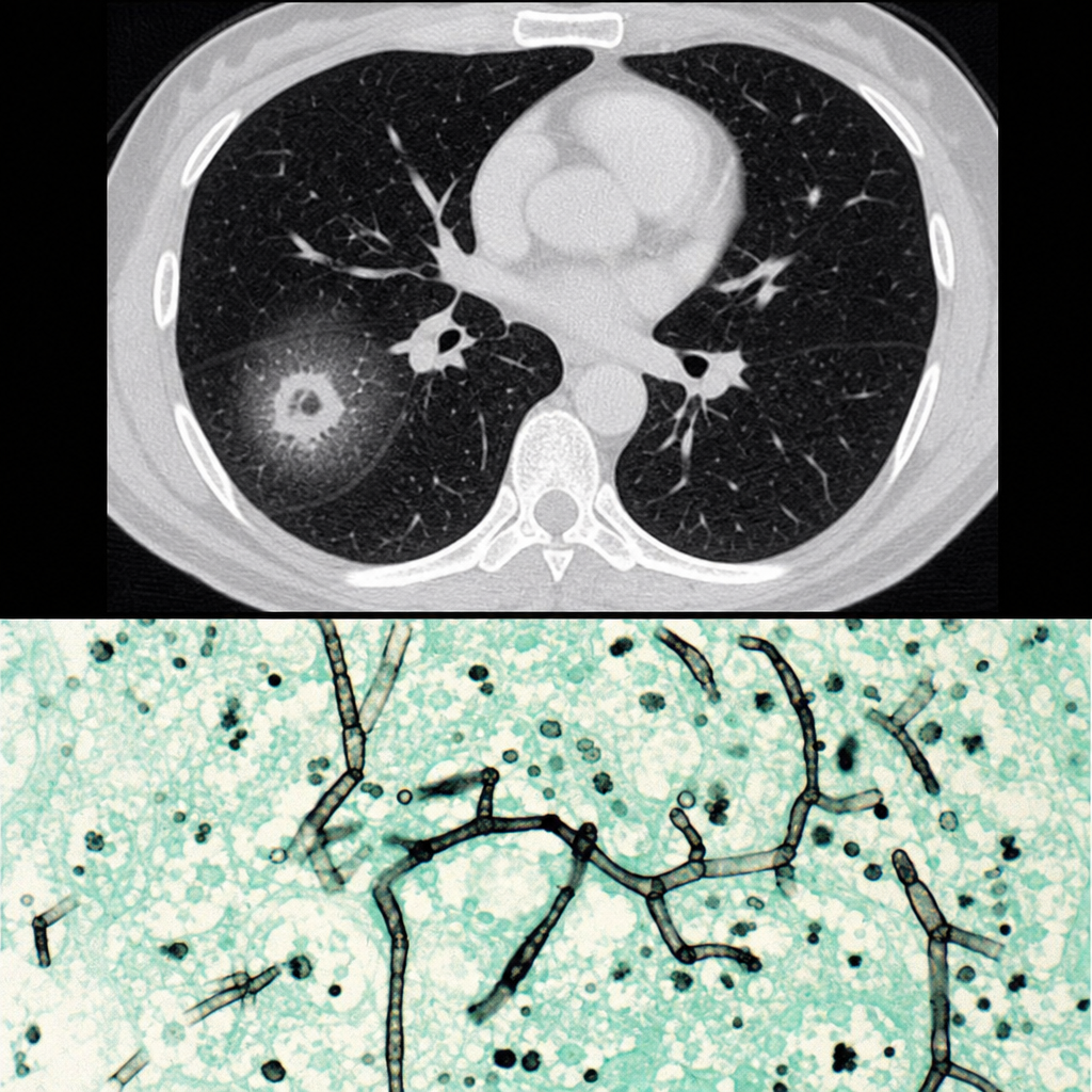
Which of the following is a non-culturable fungus?
Which of the following is not a fungal infection?
What is the most pathogenic species of Candida?
Renauld-Baudet phenomenon is seen in which of the following?
Rhinosporidium seeberi belongs to which taxonomic group?
Which organism is the most common cause of mucosal candidiasis?
Which of the following stains is used to stain fungi?
Which fungus appears as a single budding cell in culture at 37°C, where the bud and the parent yeast have a characteristic wide base of attachment?
The Raynolds-Braude phenomenon is characteristic of which organism?
Explanation: **Explanation:** *Cryptococcus neoformans* is an encapsulated yeast primarily associated with pigeon droppings and is a significant opportunistic pathogen in immunocompromised patients (e.g., HIV/AIDS). **1. Why "Urease negative" is the False Statement:** *Cryptococcus neoformans* is characteristically **Urease positive**. The production of the urease enzyme is a key biochemical marker used in the laboratory to differentiate it from other yeasts like *Candida albicans* (which is urease negative). It hydrolyzes urea to produce ammonia, raising the pH and changing the indicator color. **2. Analysis of Other Options:** * **Option A (Grows at 5°C and 37°C):** This is true. Unlike many other pathogenic fungi, *C. neoformans* can grow at 37°C (essential for human pathogenicity) and also at lower temperatures like 4°C–5°C. * **Option B (Has 4 serotypes):** This is true. Based on capsular polysaccharide antigens, it is classified into four serotypes: **A, B, C, and D**. (Note: Serotype A is *C. neoformans var. grubii*, while B and C are now often classified as *C. gattii*). * **Option D (Causes superficial skin infection):** This is true. While meningitis is the most common presentation, primary or secondary cutaneous cryptococcosis can occur, presenting as papules, pustules, or ulcerations. **High-Yield Clinical Pearls for NEET-PG:** * **Virulence Factor:** The **Polysaccharide capsule** (Glucuronoxylomannan) is the most important; it inhibits phagocytosis. * **Staining:** **India Ink** preparation shows a "halo" (negative staining). **Mucicarmine** stains the capsule red. * **Culture:** Grows on **Bird Seed Agar** (Niger Seed Agar) producing brown/black colonies due to **Phenoloxidase** activity (melanin production). * **Drug of Choice:** Induction with Amphotericin B + Flucytosine, followed by Fluconazole.
Explanation: **Explanation:** The correct answer is **Rhinosporidium seeberi**. This organism is unique in medical mycology because it has **never been successfully cultured** on artificial laboratory media (like SDA) or in cell culture. Its classification was historically debated, but molecular analysis (18S rRNA sequencing) has placed it among the *Mesomycetozoea*, a group of aquatic fish parasites, though it is still traditionally studied in Mycology. **Why the other options are incorrect:** * **Candida:** A common yeast that grows readily on Sabouraud Dextrose Agar (SDA) within 24–48 hours, forming creamy white colonies. * **Sporothrix:** A dimorphic fungus that can be cultured at 25°C (mold form with "flower-like" sporulation) and 37°C (yeast form). * **Penicillium:** A common saprophytic mold that grows rapidly in culture, characterized by its "brush-like" conidiophores. **High-Yield Clinical Pearls for NEET-PG:** * **Disease:** Rhinosporidiosis typically presents as **friable, leafy, strawberry-like polypoid masses** in the nose or nasopharynx. * **Transmission:** Associated with bathing in stagnant freshwater (ponds/tanks). * **Diagnosis:** Since it cannot be cultured, diagnosis relies on **histopathology**. Look for large **sporangia** (up to 350 µm) containing thousands of **endospores**. * **Treatment:** Surgical excision with wide-base cauterization is the treatment of choice; medical therapy (Dapsone) has limited efficacy.
Explanation: **Explanation:** The correct answer is **Actinomycosis**. Despite its name ending in "-mycosis" (a suffix typically denoting fungal infections), Actinomycosis is caused by **Actinomyces species**, which are **Gram-positive, anaerobic, non-acid-fast bacteria**. They are characterized by filamentous branching growth, which historically led to their misclassification as fungi. However, they lack a chitinous cell wall, possess a prokaryotic nucleus, and are susceptible to antibacterial agents (like Penicillin) rather than antifungals. **Analysis of Options:** * **Blastomycosis (A):** A systemic infection caused by the dimorphic fungus *Blastomyces dermatitidis*. It typically presents with pulmonary symptoms or verrucous skin lesions. * **Cryptococcus (B):** An infection caused by the encapsulated yeast *Cryptococcus neoformans*. It is a major cause of opportunistic meningitis in HIV/AIDS patients. * **Histoplasmosis (C):** A systemic fungal infection caused by *Histoplasma capsulatum*. It is a dimorphic fungus often associated with bird or bat droppings and mimics tuberculosis clinically. **High-Yield Clinical Pearls for NEET-PG:** * **Actinomyces israelii** is the most common human pathogen. It is a normal commensal of the oral cavity and female genital tract. * **Clinical Hallmark:** Presence of **"Sulfur granules"** (yellowish clumps of organisms) in the pus draining from sinus tracts. * **Classic Presentation:** "Lumpy jaw" (cervicofacial actinomycosis) following dental trauma or poor oral hygiene. * **Differential Diagnosis:** Do not confuse *Actinomyces* with *Nocardia*. Both are filamentous bacteria, but **Nocardia is aerobic and weakly acid-fast**, whereas Actinomyces is anaerobic and non-acid-fast.
Explanation: **Explanation:** **Candida albicans** is the most pathogenic and frequently isolated species of the genus *Candida*. Its high pathogenicity is attributed to several **virulence factors**, including the ability to undergo **phenotypic switching** (yeast to hyphal form), the production of extracellular hydrolytic enzymes (proteases, phospholipases), and the presence of adhesins that facilitate biofilm formation on mucosal surfaces and medical devices. **Analysis of Options:** * **C. albicans (Correct):** It is the leading cause of both superficial (oral thrush, vaginal candidiasis) and systemic candidiasis worldwide. It is uniquely identified by the **Germ Tube Test** (Reynolds-Braude phenomenon) and the production of **chlamydospores** on Cornmeal Agar. * **C. tropicalis:** A common Non-Albicans Candida (NAC), often associated with hematological malignancies and deep-seated infections, but generally less virulent than *C. albicans*. * **C. krusei:** Known for its **intrinsic resistance to Fluconazole**. While clinically significant in immunocompromised patients, it is less common and less pathogenic than *C. albicans*. * **C. stellatoidea:** Now considered a variant or sucrose-negative biotype of *C. albicans*. It is rarely isolated and primarily associated with vaginal infections. **High-Yield Clinical Pearls for NEET-PG:** * **Morphology:** *C. albicans* shows "Gram-positive" budding yeast cells with pseudohyphae. * **Culture:** Produces creamy white colonies with a characteristic "yeasty" odor on Sabouraud Dextrose Agar (SDA). * **ChromAgar:** *C. albicans* produces **light green** colored colonies. * **Drug of Choice:** Fluconazole is the standard treatment for sensitive strains, while Echinocandins (e.g., Caspofungin) are preferred for systemic infections or resistant NAC species like *C. auris*.
Explanation: **Explanation:** The **Renauld-Baudet phenomenon** refers to the characteristic microscopic appearance of *Candida albicans* when grown on specific media (like Cornmeal Agar). It describes the formation of **terminal, thick-walled, spherical resting spores known as Chlamydospores.** This is a diagnostic hallmark used in the laboratory to differentiate *C. albicans* from other *Candida* species. * **Candida albicans (Correct):** It is the primary species that exhibits the Renauld-Baudet phenomenon. When cultured under nutritionally deficient conditions (e.g., Dalmau technique on Cornmeal Agar), it produces pseudohyphae with clusters of blastoconidia and distinct, large, terminal chlamydospores. * **Candida parapsilosis (Incorrect):** While it produces pseudohyphae, they are typically "crooked" or "curved" (giant cells) and it does **not** produce terminal chlamydospores. * **Histoplasma capsulatum (Incorrect):** This is a dimorphic fungus. In its mold form, it produces tuberculate macroconidia (thick-walled with finger-like projections), not the Renauld-Baudet chlamydospores. * **Cryptococcus neoformans (Incorrect):** This is an encapsulated yeast. It does not form pseudohyphae or chlamydospores; its primary diagnostic feature is the polysaccharide capsule visualized by India Ink. **High-Yield Clinical Pearls for NEET-PG:** * **Germ Tube Test (Reynolds-Braude Phenomenon):** Often confused by name with Renauld-Baudet, this is the formation of true hyphae (no constriction at the origin) within 2 hours of incubation in serum. It is the rapid screening test for *C. albicans*. * **Culture:** On Sabouraud Dextrose Agar (SDA), *Candida* produces creamy white, smooth colonies with a characteristic "yeasty" odor. * **ChromAgar:** A differential medium where *C. albicans* appears emerald green. * **Candida dubliniensis:** The only other species that may rarely produce chlamydospores and a positive germ tube test, often seen in HIV patients.
Explanation: **Explanation:** *Rhinosporidium seeberi* is the causative agent of **Rhinosporidiosis**, a chronic granulomatous infection primarily affecting the mucous membranes of the nose and nasopharynx. **Why Option A is Correct:** Historically, *R. seeberi* was classified as a fungus due to its morphology (production of sporangia and spores) and its staining characteristics (positive for GMS, PAS, and Mucicarmine). In recent molecular phylogenetic studies (18S rRNA analysis), it has been reclassified under **Mesomycetozoea**, a group of aquatic protistan parasites located at the evolutionary boundary between animals and fungi. However, for the purpose of standard medical examinations like NEET-PG and based on traditional clinical microbiology, it is still categorized under **Mycology** as a fungus-like organism. **Why Other Options are Incorrect:** * **Option B (Bacteria):** It does not possess a prokaryotic cell wall or binary fission characteristics. * **Option C (Aquatic protistan protozoa):** While modern taxonomy places it in the *Mesomycetozoea* (DRIP clade), which are "protistan" in nature, "Protozoa" is a distinct group. In most medical exams, if "Fungus" is the intended answer based on classic textbooks, it remains the preferred choice unless the specific clade "Mesomycetozoea" is provided. * **Option D (Virus):** It is a large, multicellular eukaryotic structure (sporangium), far exceeding viral dimensions. **Clinical Pearls for NEET-PG:** * **Habitat:** Found in stagnant water and soil; common in sand harvesters and those bathing in ponds. * **Clinical Feature:** Leaf-like, friable, **strawberry-like polypoid mass** in the nose that bleeds easily (epistaxis). * **Diagnosis:** Cannot be cultured on artificial media. Diagnosis is by **histopathology** showing large **sporangia** (up to 350 µm) containing thousands of **endospores**. * **Treatment:** Surgical excision with wide cautery of the base. Dapsone is the medical adjunct of choice.
Explanation: **Explanation:** **Candida albicans** is the most common cause of both mucosal and systemic candidiasis worldwide. It is a commensal organism found in the oral cavity, gastrointestinal tract, and vagina. When the host's local or systemic immunity is compromised (e.g., HIV, diabetes, or prolonged antibiotic use), it transitions from a commensal to a pathogen. It is the primary agent responsible for oral thrush, esophageal candidiasis, and vulvovaginal candidiasis. **Analysis of Incorrect Options:** * **Candida tropicalis:** While it is a significant cause of candidemia (bloodstream infections), especially in patients with hematological malignancies and neutropenia, it is less common than *C. albicans* in mucosal lesions. * **Candida guilliermondii:** This is a rare cause of human infection, typically seen in immunocompromised patients or associated with medical devices and oncology settings. * **Candida krusei:** This species is clinically significant primarily due to its **intrinsic resistance to Fluconazole**. It is an uncommon cause of mucosal disease but a critical concern in hospital-acquired infections. **High-Yield Clinical Pearls for NEET-PG:** * **Germ Tube Test:** *C. albicans* is "Germ Tube positive" (Reynolds-Braude phenomenon). * **Morphology:** On Cornmeal Agar, *C. albicans* produces characteristic **chlamydospores**. * **Phenotypic Switching:** *C. albicans* can switch between yeast (commensal) and hyphal (pathogenic/invasive) forms. * **Drug Resistance:** While *C. albicans* is usually sensitive to azoles, **Candida glabrata** and **Candida krusei** are known for high levels of azole resistance. * **Candida auris:** An emerging multi-drug resistant (MDR) species causing global outbreaks in ICU settings.
Explanation: **Explanation:** **PAS (Periodic Acid-Schiff)** is the correct answer because it is a gold-standard histological stain for fungi. The underlying mechanism involves the oxidation of polysaccharides (specifically **chitin** and glucan) found in the fungal cell wall by periodic acid to form aldehydes. These aldehydes then react with the Schiff reagent to produce a brilliant **magenta or purplish-red color**, making the fungal elements stand out against the background. **Analysis of Incorrect Options:** * **Fontana-Masson Stain:** This is used to detect **melanin**. In mycology, it is specifically used to identify dematiaceous (pigmented) fungi or the capsule of *Cryptococcus neoformans*, but it is not a general fungal stain. * **Gomori’s Trichrome (Ferrous/Masson):** These stains are primarily used to differentiate between collagen and muscle fibers in connective tissue pathology, not for fungal identification. * **Perls’ Prussian Blue:** This is used to detect **ferric iron** (hemosiderin) in tissues. It is commonly used in cases of hemochromatosis or to identify "heart failure cells" in the lungs. **High-Yield Clinical Pearls for NEET-PG:** * **Grocott-Gomori Methenamine Silver (GMS):** The most sensitive stain for fungi; it stains them black. * **Mucicarmine:** Specifically used to identify the polysaccharide capsule of ***Cryptococcus neoformans*** (stains it bright red). * **India Ink:** A negative stain used for the rapid identification of *Cryptococcus* in CSF. * **Calcofluor White:** A fluorescent stain that binds to chitin; viewed under a UV microscope.
Explanation: **Explanation:** The question describes the classic morphology of **Blastomyces dermatitidis**, a thermally dimorphic fungus. In its parasitic yeast phase (at 37°C or in tissue), it appears as large, thick-walled, spherical cells. The defining diagnostic feature is **broad-based budding**, where the daughter cell attaches to the parent cell via a wide neck, making them appear "stuck" together. **Analysis of Options:** * **B. dermatitidis (Correct):** Known for "Broad-Based Budding" (BBB). It is primarily found in the Ohio and Mississippi River valleys and causes pulmonary infections that can disseminate to the skin and bones. * **C. neoformans:** Characterized by **narrow-based budding** and a thick polysaccharide capsule (visible with India Ink). It does not show a wide base of attachment. * **C. albicans:** Typically presents as oval budding yeast with **pseudohyphae**. At 37°C in serum, it produces **germ tubes** (true hyphae without constriction). * **P. brasiliensis:** Exhibits a "mariner’s wheel" or **"pilot’s wheel"** appearance due to multiple narrow-based buds surrounding a single large mother cell. **High-Yield Clinical Pearls for NEET-PG:** * **Mnemonic for Blastomyces:** **B**lasto = **B**ig, **B**lue (on H&E), **B**road-**B**ased **B**udding. * **Dimorphism Rule:** "Mold in the Cold (25°C), Yeast in the Heat (37°C)." * **Systemic Mycoses:** All major systemic dimorphic fungi (*Histoplasma, Blastomyces, Coccidioides, Paracoccidioides*) are acquired via inhalation of spores. * **Histoplasma capsulatum:** Small intracellular yeasts within macrophages (no broad base).
Explanation: **Explanation:** The **Reynolds-Braude phenomenon** (also known as the **Germ Tube Test**) is a rapid diagnostic test used to identify **Candida albicans**. When *C. albicans* cells are incubated in human or animal serum at 37°C for 2–3 hours, they produce elongated, tube-like projections called **germ tubes**. These are the precursors to true hyphae and are characterized by having **no constriction** at their point of origin from the parent yeast cell. This phenomenon is a key virulence factor, representing the transition from yeast to the invasive hyphal form. **Analysis of Options:** * **Candida albicans (Correct):** It is the primary medically important yeast that is germ tube positive. *Candida dubliniensis* is the only other species that typically shows a positive result. * **Mucor:** This is a Zygomycete characterized by broad, **aseptate hyphae** with right-angled (90°) branching. It does not produce germ tubes. * **Cryptococcus:** This is an encapsulated yeast. It is identified by **India Ink** preparation (showing a halo) and the **Urease test**, but it does not form germ tubes. * **Aspergillus:** This is a filamentous fungus (mould) characterized by septate hyphae with **acute-angled (45°) branching**. It reproduces via conidia, not germ tube formation from yeast cells. **High-Yield Clinical Pearls for NEET-PG:** * **Culture Media:** *C. albicans* produces "Creamy white" colonies with a characteristic "yeasty" odor on **Sabouraud Dextrose Agar (SDA)**. * **Chlamydospores:** On **Cornmeal Agar**, *C. albicans* produces thick-walled terminal chlamydospores. * **Morphology:** It is a dimorphic fungus (technically polymorphic) existing as yeast, pseudohyphae, and true hyphae. * **Drug of Choice:** Fluconazole for localized infections; Echinocandins (like Caspofungin) or Amphotericin B for systemic candidiasis.
Classification of Fungi
Practice Questions
Superficial Mycoses
Practice Questions
Dermatophytes
Practice Questions
Subcutaneous Mycoses
Practice Questions
Candidiasis
Practice Questions
Aspergillosis
Practice Questions
Cryptococcosis
Practice Questions
Zygomycosis
Practice Questions
Endemic Mycoses
Practice Questions
Opportunistic Fungal Infections
Practice Questions
Antifungal Agents
Practice Questions
Laboratory Diagnosis of Fungal Infections
Practice Questions
Get full access to all questions, explanations, and performance tracking.
Start For Free