Which one of the following is an emerging viral disease?
What is the best investigation for identifying malaria species?
Which of the following techniques is primarily used for RNA analysis?
Best method for the detection of mutations with low allele frequency is:
All of the following are pattern recognition receptors for extracellular or ingested microbes, except:
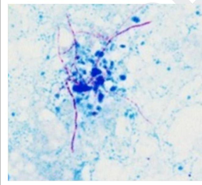
Gene amplification is achieved through
DNA amplification is done by all, except:
Influenza virus causes new epidemic by (3-5 yrs)-
What does Polymerase Chain Reaction (PCR) detect?
Acridine orange is a fluorescent dye used to bind which cellular components?
Explanation: ***SARS*** - **SARS (Severe Acute Respiratory Syndrome)** emerged in 2002-2003 and caused a global outbreak, making it a classic example of an **emerging viral disease**. - Emerging viral diseases are those that have recently appeared in a population or whose incidence has increased rapidly in recent times. *Measles* - Measles is an **established and re-emerging disease**, meaning it has been present for a long time but has seen resurgence due to factors like declining vaccination rates. - It is caused by the **measles virus (MeV)**, a paramyxovirus, and has been a known human pathogen for centuries. *Chicken pox* - Chickenpox, caused by the **varicella-zoster virus (VZV)**, is a common and well-known childhood disease that has been endemic for a long time. - While it can be severe, it is not considered an emerging disease as its presence and characteristics have been established for many years. *Rabies* - Rabies, caused by the **rabies virus**, is a highly fatal disease primarily transmitted through animal bites and has been recognized globally for centuries. - Although it remains a significant public health concern, it is an **ancient and well-established zoonotic disease**, not an emerging one.
Explanation: ***Thin smear with Giemsa*** - A **thin smear** allows for the visualization of **parasite morphology** within red blood cells, which is crucial for distinguishing between species of *Plasmodium*. - **Giemsa stain** provides optimal contrast for identifying characteristic features such as **merozoites**, **trophozoites**, **schizonts**, and **gametocytes** of different malaria species. *Thick smear* - A **thick smear** is primarily used for **detecting the presence of malaria parasites** and for quantifying parasite density due to its higher sensitivity. - However, because red blood cells are lysed, it **does not preserve parasite morphology** well, making species identification difficult. *QBC* - **Quantitative Buffy Coat (QBC) analysis** is a rapid method for detecting malaria parasites based on their fluorescence under UV light. - While sensitive for detection, it generally **does not allow for precise species identification** due to the lack of clear morphological detail. *Thin smear with acridine orange* - A **thin smear stained with acridine orange** is used for rapid detection of parasites by fluorescence microscopy. - Similar to QBC, it is **less effective for detailed morphological examination** and specific species identification compared to Giemsa-stained thin smears.
Explanation: ***Next generation sequencing*** - **Next-generation sequencing (NGS)**, particularly RNA-Seq, is widely used for **transcriptome analysis** to quantify and discover RNA molecules. - RNA-Seq allows for the precise measurement of **gene expression levels**, identification of **novel transcripts**, and detection of **splicing variants**. *Sanger's technique* - **Sanger sequencing** is primarily used for **DNA sequencing** to determine the exact order of nucleotides in a DNA molecule. - While it can be applied to cDNA (synthesized from RNA), it is not directly used for **RNA analysis** itself. *Western blot* - **Western blot** is a laboratory technique used to detect specific **proteins** in a sample. - It involves separating proteins by size using gel electrophoresis and then transferring them to a membrane for antibody-based detection, making it unsuitable for direct **RNA analysis**. *PCR* - **Polymerase Chain Reaction (PCR)** is used to amplify specific **DNA sequences**. - While **Reverse Transcription PCR (RT-PCR)** can quantify RNA by first converting it to cDNA, PCR itself does not directly analyze the RNA molecule.
Explanation: ***Droplet digital PCR*** - **Droplet digital PCR (ddPCR)** offers superior sensitivity for detecting **low allele frequency mutations** by partitioning the sample into thousands of individual reactions. - This compartmentalization allows for the direct quantification of target DNA molecules without relying on a standard curve, making it highly accurate for rare mutation detection. *FISH* - **Fluorescence in situ hybridization (FISH)** primarily detects **chromosomal abnormalities** like translocations, deletions, or amplifications, rather than single-nucleotide variants or small indels with low allele frequencies [2]. - It visualizes genetic changes at a **cytogenetic level** on an intracellular basis, not typically for quantifying rare DNA mutations in a heterogeneous sample. *Sanger sequencing* - **Sanger sequencing** is the gold standard for **sequencing individual DNA fragments** but has a detection limit of around 15-20% for allele frequency, making it unsuitable for very low allele frequency mutations [1]. - It struggles to reliably detect minor alleles when they are present in a small proportion of the total DNA pool. *Nested PCR* - **Nested PCR** increases the sensitivity and specificity of amplification by using two sets of primers in a sequential manner but does not inherently provide the **quantification capability** or the same level of **low allele frequency detection** as ddPCR processes. - While sensitive for detecting target sequences, it is not designed for precise quantification of rare mutations in a background of wild-type sequences. **References:** [1] Kumar V, Abbas AK, et al.. Robbins and Cotran Pathologic Basis of Disease. 9th ed. Genetic Disorders, p. 185. [2] Kumar V, Abbas AK, et al.. Robbins and Cotran Pathologic Basis of Disease. 9th ed. Genetic Disorders, pp. 185-186.
Explanation: ***Killer-cell immunoglobulin receptors (KIRs)*** - KIRs are primarily involved in regulating **natural killer (NK) cells**, not in recognizing microbes [1]. - They primarily interact with **MHC class I molecules** and play a role in **immune surveillance**, rather than pattern recognition of pathogens [1]. *NOD-like receptors (NLRs)* - NLRs detect **intracellular pathogens** and damaged cells, playing a crucial role in **innate immunity** [2]. - They initiate responses to **bacterial peptidoglycans** and work in the recognition of microbial patterns [2]. *Toll-like receptors (TLRs)* - TLRs are well-known for recognizing **extracellular microbes** and activate the immune response upon pathogen detection [3]. - They are critical in detecting **lipopolysaccharides (LPS)** and **viral nucleic acids** to elicit immune responses. *C-type lectin receptors (CLRs)* - CLRs specifically recognize **carbohydrate structures** on pathogens, playing a key role in **innate immune responses** [3]. - They are important in identifying **fungi** and **bacteria**, enhancing phagocytosis and cytokine production. **References:** [1] Kumar V, Abbas AK, et al.. Robbins and Cotran Pathologic Basis of Disease. 9th ed. Diseases of the Immune System, pp. 200-201. [2] Kumar V, Abbas AK, et al.. Robbins and Cotran Pathologic Basis of Disease. 9th ed. Diseases of the Immune System, p. 196. [3] Kumar V, Abbas AK, et al.. Robbins and Cotran Pathologic Basis of Disease. 9th ed. Diseases of the Immune System, p. 200.
Explanation: ***Polymerase Chain Reaction*** - **PCR** is the **gold standard** molecular biology technique that generates **millions to billions of copies** of a specific DNA segment over a short period. - It utilizes a cyclical process of **denaturation**, **annealing**, and **extension** with **thermostable DNA polymerase** to achieve exponential amplification. - **Most widely used** method for gene amplification in research and diagnostics. *DNA strand hybridization* - **DNA strand hybridization** is the process where two complementary single-stranded DNA molecules bind together to form a **double-stranded molecule**. - This process is fundamental to many molecular techniques but does not, in itself, achieve **amplification**; rather, it is a **binding event**. *In situ DNA hybridization* - **In situ hybridization** is a technique that localizes and detects specific **nucleic acid sequences** (DNA or RNA) within cells or tissues directly on a slide. - While it uses **hybridization**, its primary purpose is **detection and localization**, not the **amplification** of DNA sequences. *Ligase chain reaction (LCR)* - **LCR** is a molecular technique that does amplify DNA sequences exponentially using **DNA ligase** to join adjacent oligonucleotide probes. - However, it is **less commonly used** than PCR, has more **stringent requirements** (requires knowledge of both strands), and is primarily used for detecting **known point mutations** rather than general gene amplification. - **PCR remains the standard** technique when the question refers to gene amplification without additional qualifiers.
Explanation: ***DNA sequencing*** - **DNA sequencing** determines the **nucleotide base order** in a DNA molecule but does not increase the amount of DNA. - While requiring a DNA template, it is an **analytical technique** rather than an amplification method. *Loop-mediated isothermal amplification (LAMP)* - **LAMP** is an **isothermal DNA amplification** technique that amplifies target DNA sequences at a constant temperature (60-65°C). - It uses a DNA polymerase with strand displacement activity and 4-6 primers to produce large amounts of DNA rapidly. *Ligase chain reaction* - **LCR** is an amplification method that detects specific **DNA sequences** by ligating adjacent probes. - It amplifies the signal from a target DNA sequence rather than the DNA itself by creating many copies of joined probes. *Polymerase chain reaction* - **PCR** is a widely used technique for **amplifying** a specific segment of DNA to produce many copies. - It involves cycles of **denaturation**, **annealing**, and **extension** using a DNA polymerase.
Explanation: ***Antigenic drift*** - **Antigenic drift** involves minor changes in the **hemagglutinin (HA)** and **neuraminidase (NA)** surface proteins of the influenza virus due to point mutations. - These minor changes allow the virus to slightly evade the host's immune system, leading to **seasonal epidemics** (typically every 2-3 years) as pre-existing immunity is less effective. *Cycle trends* - This term is too general and does not specifically describe the ** virological mechanism** responsible for influenza epidemics. - While influenza does exhibit cyclical patterns, "cycle trends" doesn't explain the underlying biological process of viral evolution. *Mosaicism* - **Mosaicism** refers to the presence of two or more populations of cells with different genotypes within a single individual. - This genetic phenomenon is completely unrelated to how influenza viruses cause new epidemics. *Antigenic shift* - **Antigenic shift** involves abrupt, major changes in the HA or NA proteins, usually through **gene reassortment** when two different influenza viruses co-infect the same cell. - This leads to entirely **new viral subtypes** that can cause global **pandemics** (less frequently, perhaps every 10-40 years), not the more regular 3-5 year epidemics.
Explanation: **Explanation:** **Why Nucleic Acid is the Correct Answer:** Polymerase Chain Reaction (PCR) is a molecular technique used to **amplify specific sequences of DNA**. It utilizes a heat-stable DNA polymerase (like *Taq* polymerase) to create millions of copies of a target genetic sequence. In microbiology, PCR is used to detect the **nucleic acid** (DNA or RNA) of a pathogen. For RNA viruses (like HIV or SARS-CoV-2), a variation called Reverse Transcription-PCR (RT-PCR) is used to first convert RNA into complementary DNA (cDNA) before amplification. **Why Other Options are Incorrect:** * **Antigens (Option A):** These are proteins or polysaccharides on the surface of a pathogen. They are detected using immunological assays like **ELISA** (Enzyme-Linked Immunosorbent Assay) or Lateral Flow Assays (Rapid Antigen Tests), not PCR. * **Antibodies (Option B):** These are host proteins produced by B-cells in response to an infection. They are detected via **Serology** (e.g., ELISA, Western Blot, or Agglutination tests) to identify past or current exposure, whereas PCR identifies the presence of the organism itself. **High-Yield Clinical Pearls for NEET-PG:** * **Steps of PCR:** Denaturation (94-96°C) → Annealing (50-65°C) → Extension (72°C). * **Real-Time PCR (qPCR):** Allows for **quantification** of the microbial load (e.g., Viral Load in Hepatitis C or HIV). * **Multiplex PCR:** Can detect multiple different pathogens in a single clinical sample simultaneously using different primers. * **Sensitivity:** PCR is highly sensitive, making it the "Gold Standard" for diagnosing organisms that are difficult to culture (e.g., *M. tuberculosis*, *Chlamydia*, or viral infections).
Explanation: **Explanation:** **Acridine orange** is a fluorochrome dye that functions as a nucleic acid-selective stain. It has the unique property of **metachromasia**, meaning it can differentiate between double-stranded and single-stranded nucleic acids based on the wavelength of light emitted. 1. **Why A is Correct:** Acridine orange intercalates into **DNA** (double-stranded) and binds electrostatically to **RNA** (single-stranded). When excited by blue light (460 nm) under a fluorescence microscope, DNA-bound dye emits **green fluorescence**, while RNA-bound dye emits **orange-red fluorescence**. This makes it highly effective for detecting microorganisms in clinical specimens (like blood cultures or CSF) where bacteria/fungi appear bright against a dark background. 2. **Why Other Options are Incorrect:** * **B (Proteins):** Proteins are typically stained with dyes like Coomassie Brilliant Blue or Silver stain. * **C (Lipids):** Lipids are visualized using lipophilic stains such as Sudan Black or Oil Red O. * **D (Carbohydrates):** Carbohydrates (glycogen/mucin) are identified using the Periodic Acid-Schiff (PAS) stain. **High-Yield Clinical Pearls for NEET-PG:** * **Sensitivity:** Acridine orange is more sensitive than the Gram stain for detecting low concentrations of bacteria (e.g., in buffy coat smears or early positive blood cultures). * **Rapid Screening:** It is used for rapid screening of malaria parasites (QBC technique) and *Trichomonas vaginalis*. * **Cell Viability:** It can distinguish between live (green) and dead (red/orange) cells in certain laboratory assays.
Get full access to all questions, explanations, and performance tracking.
Start For Free