What is the diagnosis of an umbilicated, pearly white, asymptomatic skin lesion?
A diabetic patient developed cellulitis due to S. aureus, which was found to be methicillin resistant on the antibiotic sensitivity testing. All of the following antibiotics will be appropriate except ?
A diabetic patient presents with sudden-onset perineal pain. On examination, foul-smelling discharge and necrotic tissue are noted. Which of the following is the most characteristic feature of this condition?
A 50-year-old diabetic presents with a foot ulcer. Which pathogen is most likely?
What is the first-line treatment for gas gangrene?
Which of the following conditions is caused by Staphylococcus aureus?
Cellulitis is characterized as:
A child presents with grouped vesicles on the lips. What is the bedside investigation that you would like to do?
A 50-year-old diabetic presents with a foot ulcer. Which pathogen is most likely?
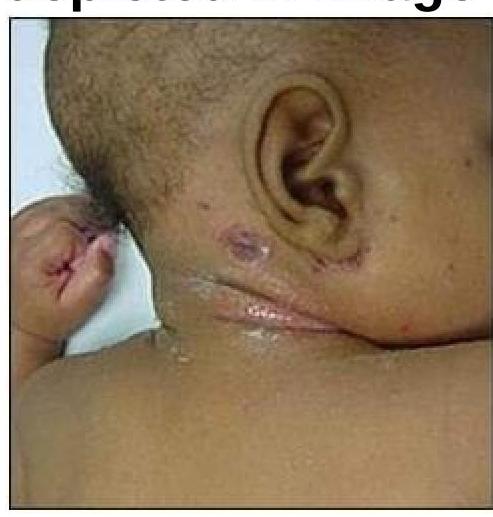
What is the causative organism for the condition depicted in the image?

Explanation: ***Molluscum contagiosum*** - This **viral skin infection** typically presents with **multiple, small (2-5 mm), firm, pearly, dome-shaped papules** that have a **central umbilication**. - The lesions are usually **asymptomatic**, as described, though they can occasionally be itchy or inflamed. - Caused by a **poxvirus** and is highly contagious through direct contact. *EBV* - **Epstein-Barr Virus (EBV)** is primarily associated with **infectious mononucleosis**, which presents with fever, sore throat, and lymphadenopathy, not umbilicated skin lesions. - EBV can cause oral hairy leukoplakia in immunocompromised individuals, which is a white lesion, but it is **not pearly, umbilicated, or dome-shaped**. *HSV* - **Herpes Simplex Virus (HSV)** causes lesions that are typically **grouped vesicles on an erythematous base** that evolve into erosions or ulcers. - HSV lesions are often **painful or itchy** and **do not appear as pearly, umbilicated papules**. *None of the options* - This is incorrect because **Molluscum contagiosum** perfectly matches the clinical description of umbilicated, pearly white, asymptomatic skin lesions. - The classic **central umbilication** is the pathognomonic feature that distinguishes molluscum from other viral skin infections.
Explanation: ***Imipenem*** - **Imipenem** is a carbapenem antibiotic that is effective against many Gram-positive and Gram-negative bacteria, but it is **not active against MRSA (methicillin-resistant *Staphylococcus aureus*)**. - MRSA strains are resistant to all beta-lactam antibiotics, including penicillins, cephalosporins, and carbapenems like imipenem, due to the presence of the **mecA gene** which encodes for an altered penicillin-binding protein (PBP2a). *Vancomycin* - **Vancomycin** is a glycopeptide antibiotic that is a primary choice for treating **MRSA infections**, including cellulitis. - It inhibits cell wall synthesis by binding to the D-Ala-D-Ala precursor, preventing cross-linking, and is specifically active against **Gram-positive bacteria**. *Teicoplanin* - **Teicoplanin** is another glycopeptide antibiotic, similar to vancomycin, and is also considered a suitable agent for treating **MRSA infections**. - It works by inhibiting bacterial cell wall synthesis and has a **longer half-life** than vancomycin, allowing for less frequent dosing. *Linezolid* - **Linezolid** is an oxazolidinone antibiotic known for its activity against **Gram-positive bacteria**, including **MRSA** and vancomycin-resistant enterococci (VRE). - It inhibits protein synthesis by binding to the 50S ribosomal subunit, preventing the formation of the initiation complex.
Explanation: **Mixed aerobic and anaerobic infection** - Fournier's gangrene is a polymicrobial infection typically involving a **synergistic mixture of aerobic and anaerobic bacteria**. - This mixed infection contributes to the rapid progression and tissue destruction seen in this condition, leading to the **foul-smelling discharge** due to anaerobic metabolism. *Anti-gas gangrene serum is indicated only in specific cases.* - Anti-gas gangrene serum is specifically for **Clostridium perfringens** infections, which can cause gas gangrene but is usually a distinct clinical entity from Fournier's. - While Clostridium species can be present in Fournier's gangrene, it is not the sole causative agent, and **broader antimicrobial therapy** is the mainstay of treatment, not antitoxin serum. *Urinary diversion may be considered in severe cases.* - Urinary diversion, such as a **suprapubic catheter**, may be necessary when the urethra or perineum is extensively involved or to prevent ongoing contamination of the surgical site. - However, it's not a primary treatment for the infection itself but rather an **adjunctive measure** to manage complicated cases of Fournier's gangrene. *Bilateral orchidectomy is not routinely required.* - **Testicular involvement** in Fournier's gangrene is rare due to the separate blood supply of the testes. - **Orchidectomy** is only performed if the testes themselves are affected by necrosis, which is uncommon and occurs in critically severe cases; routine removal is not indicated.
Explanation: ***Staphylococcus aureus*** - **_Staphylococcus aureus_** is the most common pathogen isolated from **diabetic foot ulcers** due to its prevalence on the skin and ability to infect compromised tissues. - Diabetic patients are particularly susceptible to **_S. aureus_** infections due to **impaired immune function** and **poor circulation**. *Pseudomonas aeruginosa* - While _Pseudomonas aeruginosa_ can cause foot infections, it is typically associated with **chronic, wet wounds** or those exposed to water, and is less common as a primary pathogen than _S. aureus_. - Infections by _Pseudomonas_ often result in a **greenish discharge** and a characteristic fruity odor, which are not mentioned here. *Escherichia coli* - **_Escherichia coli_** is primarily a cause of **urinary tract infections** and **gastrointestinal infections**. - While it can be found in polymicrobial wound infections, it is not the most likely single pathogen to initiate a diabetic foot ulcer infection. *Candida albicans* - **_Candida albicans_** is a **fungus** that can cause infections, particularly in immunocompromised individuals and in moist areas. - While **fungal infections** can complicate diabetic foot ulcers, it is not the primary bacterial pathogen typically responsible for the initial presentation of such ulcers.
Explanation: ***Debridement & antibiotics*** - **Aggressive surgical debridement** to remove necrotic tissue and reduce bacterial load is the most critical initial step. - **Broad-spectrum antibiotics**, particularly penicillin G, are essential to target the causative *Clostridium perfringens* and prevent systemic spread. *Hyperbaric oxygen* - While **hyperbaric oxygen therapy** can be a useful adjunct by inhibiting bacterial growth and toxin production in anaerobic environments, it is not the *first-line* or sole treatment. - It should be used in conjunction with debridement and antibiotics, not as a standalone initial therapy. *Polyvalent gas gangrene antitoxin* - **Antitoxins** are generally not recommended due to their limited efficacy and potential for severe allergic reactions. - The primary treatment focuses on removing the source of infection and killing the bacteria, not neutralizing toxins alone. *Amputation* - **Amputation** is a drastic measure typically reserved for cases where the limb is irreversibly damaged, infection is uncontrollable by other means, or there is a threat to life. - It is not the initial treatment but may be necessary in advanced or complicated cases.
Explanation: ***Bullous impetigo*** - Bullous impetigo is a superficial skin infection characterized by **blisters (bullae)**, and is specifically caused by **Staphylococcus aureus** producing exfoliative toxins. - The toxins produced by *S. aureus* cause intraepidermal cleavage, leading to the formation of the characteristic **flaccid bullae**. *Corynebacterium minutissimum infection* - *Corynebacterium minutissimum* causes **erythrasma**, a chronic superficial skin infection characterized by well-demarcated reddish-brown patches, often in intertriginous areas. - It does not cause bullous impetigo and is typically diagnosed by its coral-red fluorescence under a **Wood's lamp**. *Haemophilus ducreyi infection* - *Haemophilus ducreyi* is the causative agent of **chancroid**, a sexually transmitted infection characterized by painful genital ulcers with a necrotic base and often accompanied by swollen, tender regional lymph nodes. - It is not associated with skin blistering or bullous impetigo. *Propionibacterium acnes infection* - *Propionibacterium acnes* (now *Cutibacterium acnes*) is a bacterium commonly implicated in **acne vulgaris**, contributing to inflammation and comedone formation within hair follicles. - It causes inflammatory lesions like papules, pustules, nodules, and cysts, rather than bullous lesions.
Explanation: ***Nonsuppurative and invasive*** - Cellulitis is considered **nonsuppurative** as it typically lacks macroscopic pus formation, distinguishing it from abscesses. - It is **invasive** because it involves the dermal and subcutaneous tissues, spreading through fascial planes. *Suppurative and invasive* - This description is more indicative of conditions like an **abscess**, which involves localized collections of pus. - While abscesses are invasive, cellulitis characteristically lacks the discrete pus collection. *Nonsuppurative and non-invasive* - Conditions that are nonsuppurative and non-invasive might include self-limiting skin rashes or superficial inflammatory processes. - Cellulitis involves deeper tissue infection, which inherently makes it invasive. *Suppurative and non-invasive* - A condition that is suppurative but non-invasive would be rare and contradictory, as pus formation often indicates a tissue response that is at least locally invasive. - Superficial pustules might be considered suppurative and relatively non-invasive, but cellulitis clearly extends beyond such superficial lesions.
Explanation: ***Tzanck smear*** - A **Tzanck smear** is a rapid bedside test that can identify **multinucleated giant cells**, which are seen in herpes simplex virus infections. - The presence of **grouped vesicles on the lips** is highly suggestive of **herpes labialis** (HSV-1), which is primarily a **clinical diagnosis**. - Among the options provided, Tzanck smear is the only relevant bedside investigation, though it has **limited sensitivity and specificity** and **cannot distinguish between HSV and VZV**. - In modern practice, **PCR or direct immunofluorescence** are preferred when laboratory confirmation is needed, but Tzanck smear remains a low-cost option in resource-limited settings. *Wood's lamp* - A Wood's lamp uses **ultraviolet light** to detect certain fungal or bacterial infections by revealing characteristic fluorescence. - It is useful for conditions like **tinea capitis** (green fluorescence) and **erythrasma** (coral-red fluorescence), but has no role in diagnosing viral vesicular lesions. *Slit skin smear* - A **slit skin smear** is used to detect **acid-fast bacilli** in the diagnosis of **leprosy**. - It is not indicated for vesicular lesions and is irrelevant to herpes simplex infection. *KOH* - A **KOH (potassium hydroxide) mount** is used to diagnose **fungal infections** by dissolving keratinocytes and revealing fungal hyphae or spores. - It has no utility in diagnosing viral infections such as herpes simplex.
Explanation: ***Staphylococcus aureus*** - **Staphylococcus aureus** is the most common bacterial pathogen isolated from **diabetic foot ulcers**, often due to compromised skin integrity and neuropathy. - It can cause a range of infections, from superficial cellulitis to deep tissue infections and osteomyelitis, common in diabetic foot. *Pseudomonas aeruginosa* - While *Pseudomonas aeruginosa* can infect foot ulcers, especially in patients with **previous antibiotic exposure** or **immersion injuries**, it is less common as a primary pathogen than *S. aureus*. - Infections by *Pseudomonas* often present with a **characteristic sweet, grape-like odor** and a green-blue exudate. *Escherichia coli* - *Escherichia coli* is generally associated with infections originating from the **gastrointestinal or genitourinary tracts** and is not a typical primary cause of foot ulcers. - Its presence in foot ulcers is rare and often suggests **polymicrobial infection** or fecal contamination. *Candida albicans* - *Candida albicans* is a **fungal pathogen** that can cause infections, particularly in immunocompromised individuals or those with chronic moist conditions between the toes. - While diabetics are prone to **fungal infections**, *Candida* is not a common primary cause of a deep foot ulcer; bacterial infections are far more prevalent.
Explanation: ***Staphylococci*** - The image shows **impetigo** with **crusted lesions**, consistent with **_Staphylococcus aureus_** infection. - **Staphylococcus aureus** is a major causative organism of impetigo, particularly **bullous impetigo**, and commonly produces the characteristic **honey-colored crusts** seen in non-bullous forms as well. - This superficial bacterial skin infection is highly contagious and responds well to topical or systemic antibiotics. *Candidal infection* - **Candidal infections** (e.g., candidiasis) typically present as **erythematous patches** with satellite lesions, or white plaques in mucosal areas, which is not consistent with the image. - This fungal infection is often seen in immunocompromised individuals or in warm, moist skin folds, not as crusted superficial lesions. *Streptococcus* - While **_Streptococcus pyogenes_** can also cause impetigo (especially non-bullous impetigo), the clinical presentation in the image is most consistent with **staphylococcal infection**. - Streptococcal infections may present similarly but can also cause other conditions like cellulitis or erysipelas with distinct features. *Actinomycetes* - **Actinomycosis** is a rare, chronic bacterial infection that forms **abscesses and sinus tracts**, often with "sulfur granules," which is distinct from the superficial skin lesions shown. - This infection usually involves deeper tissues and presents as a chronic, indolent infection, unlike the acute superficial presentation of impetigo.
Get full access to all questions, explanations, and performance tracking.
Start For Free