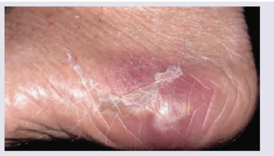
A patient walking barefoot during his morning walk has developed a swelling in the foot. What is the probable diagnosis?

Which of the following organisms is incriminated in a patient of left sided endocarditis involving the mitral valve? (Recent NEET Pattern 2016-17)
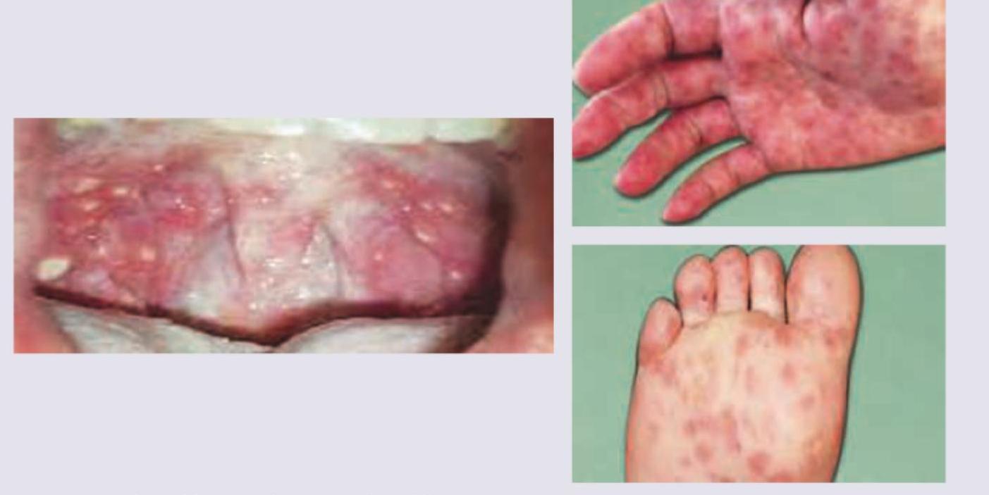
All are correct about the condition shown in the image except:

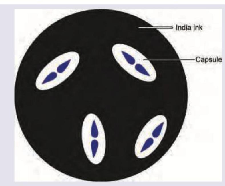
A 25-year-old truck driver presents with history of fever for 3 days with altered sensorium for 1 day. On the way to hospital he had an episode of vomiting followed by seizures. On examination reflexes are brisk and neck stiffness was noted. Mannitol was given and Lumbar puncture was performed. The microscopic examination of CSF sample yields the view given below. What is the diagnosis?

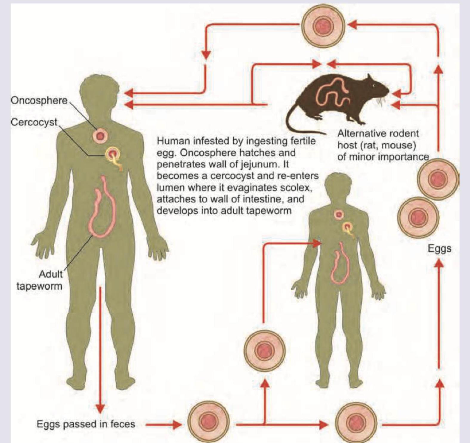
Which of the following life cycles is shown below?

Which causative organism is responsible for this disease?

All of the following organisms are involved in post-splenectomy sepsis except
What is the mechanism behind the increased risk of HIV acquisition in individuals with genital herpes?
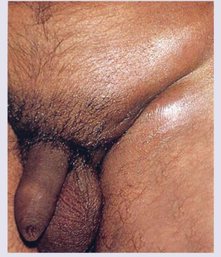
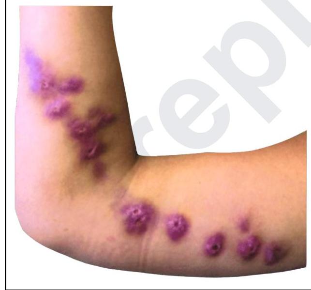
A young man presents with skin lesions as shown in the image below. All of the following organisms can spread through dermal and subcutaneous lymphatics, except

A 34-year-old man comes to the physician because of fatigue and shortness of breath with moderate exertion for the past 2 months. Over the past 10 days, he has had low-grade fevers and night sweats. He has no history of serious illness except for a bicuspid aortic valve diagnosed 5 years ago. He has smoked one pack of cigarettes daily for 10 years and drinks 3–5 beers on social occasions. He does not use illicit drugs. The patient takes no medications. He appears weak. His temperature is 37.7°C (99.9°F), pulse is 70/min, and blood pressure is 128/64 mm Hg. The lungs are clear to auscultation. A grade 2/6 systolic murmur is heard best at the right sternal border and second intercostal space. There are several hemorrhages underneath his fingernails on both hands and multiple tender, red nodules on his fingers. Which of the following is the most likely causal organism?
Explanation: **Madura foot (Correct Answer)** - **Madura foot**, or mycetoma, is a **chronic granulomatous infection** often acquired through minor skin trauma, such as walking barefoot on contaminated soil - Classic presentation: **localized swelling, draining sinuses, and grain formation** - The image shows a **swollen foot** with signs of chronic infection, consistent with the progressive nature of Madura foot affecting subcutaneous tissues and eventually bone - Endemic in tropical regions including India, making this the most likely diagnosis given barefoot walking *Staphylococcus aureus (Incorrect)* - While *S. aureus* can cause various skin infections and swelling, it typically presents with **acute infections** (abscesses, cellulitis, folliculitis) - A **chronic, localized swelling with potential sinus tracts** that progresses over time is less typical for uncomplicated *S. aureus* infections - The context of barefoot walking on soil is more suggestive of fungal or actinomycotic infections *Botryomycosis (Incorrect)* - **Botryomycosis** is a rare chronic bacterial infection that causes granulomas and abscesses, often with "grains" similar to mycetoma - Typically caused by bacteria like *Staphylococcus aureus* or *Pseudomonas aeruginosa*, **not typically acquired directly from soil** - Though it presents with granulomas and "grains," the context of walking barefoot and the endemic nature of mycetoma in India makes Madura foot more likely *Tetanus (Incorrect)* - **Tetanus** is a severe neurological condition caused by the toxin of *Clostridium tetani*, entering through wounds - Presents with **muscle spasms, rigidity, and lockjaw** (trismus) - Does **not cause localized swelling or chronic granulomatous lesions** as shown in the image - Wrong clinical presentation entirely
Explanation: ***Streptococcus viridans*** - This group of bacteria is the **most common cause** of **subacute infective endocarditis** on native, previously damaged valves, particularly the mitral valve. - They typically colonize the oral cavity and can enter the bloodstream after dental procedures or poor oral hygiene, leading to seeding of cardiac valves. *Candida albicans* - *Candida albicans* is a common cause of **fungal endocarditis**, which typically presents as a more subacute or chronic illness and can occur in immunocompromised individuals or those with indwelling catheters. - While it can affect the mitral valve, it is less common than bacterial causes, especially in the context of left-sided endocarditis unless specific risk factors (e.g., intravenous drug use, prosthetic valves, prolonged antibiotic use) are present. *Enterococci* - **Enterococcal endocarditis** often occurs in older patients, those with underlying genitourinary or gastrointestinal tract pathology, or healthcare-associated infections. - While they can affect native or prosthetic valves, they are not the most common cause of native valve endocarditis involving the mitral valve in the general population. *Pseudomonas* - **Pseudomonas endocarditis** is typically associated with **intravenous drug use** and commonly affects the **tricuspid valve** (right-sided endocarditis). - While it can involve left-sided valves, it is less frequent in the absence of intravenous drug use compared to *Streptococcus viridans*.
Explanation: ***Correct: Aspirated material shows LCL bodies*** - **"LCL bodies" is NOT a recognized diagnostic term** for lymphogranuloma venereum (LGV) - The aspirated material from buboes in LGV shows **Chlamydia trachomatis elementary bodies and reticulate bodies**, along with inflammatory cells - This is the EXCEPTION as it is an incorrect statement about LGV *Incorrect: Groove sign of LGV* - The **groove sign is a classic clinical feature** of LGV - Formed by enlarged inguinal lymph nodes above and below the inguinal (Poupart's) ligament, creating a characteristic depression - This is a TRUE statement about LGV *Incorrect: Most common cause of this infection is L2 biovar* - LGV is caused by **Chlamydia trachomatis serovars L1, L2, and L3** - **L2 serovar is the most prevalent cause** of LGV infections worldwide - This is a TRUE statement about LGV *Incorrect: Females develop esthiomene* - **Esthiomene is a late complication** of chronic untreated LGV in females - Characterized by progressive genital elephantiasis, ulceration, and destruction of vulvo-perineal tissues - Results from persistent lymphatic obstruction and chronic inflammation - This is a TRUE statement about LGV
Explanation: ***Cryptococcus*** - The image provided depicts an **India ink stain** of CSF, which is characteristic for visualizing the large polysaccharide **capsule** of *Cryptococcus neoformans* as a **clear halo** against a dark background. - Clinical features of fever, altered sensorium, seizures, brisk reflexes, and **neck stiffness** are consistent with meningitis, and *Cryptococcus* is a common cause, particularly in immunocompromised individuals or those with environmental exposure (like a truck driver who might be exposed to bird droppings). *Pneumococcus* - *Streptococcus pneumoniae* (Pneumococcus) is a common cause of bacterial meningitis, but it is a **bacterium, not a fungus**, and would not show a distinct capsule with India ink stain. - Microscopic examination of CSF for Pneumococcus would typically reveal **Gram-positive diplococci**, not encapsulated yeast forms. *Enterococcus* - *Enterococcus* species are **Gram-positive cocci** that can cause meningitis, especially in neonates, the elderly, or hospitalized patients, but they are also **bacteria**. - They also would not present with a capsule visible by India ink staining; routine Gram stain would be used for their identification. *Listeria monocytogenes* - *Listeria monocytogenes* is a **Gram-positive rod** that causes meningitis, particularly in pregnant women, neonates, the elderly, and immunocompromised individuals. - Similar to the other bacterial options, it is identified by **Gram stain** and culture, and an India ink stain would not reveal encapsulated yeast.
Explanation: ***Hymenolepis nana*** - The image clearly depicts the life cycle of *Hymenolepis nana*, showing direct human infection from ingesting **fertile eggs**, leading to the development of an **oncosphere** and then a **cercocyst** within the human host before maturing into an adult tapeworm in the intestine. - The presence of an "alternative rodent host (rat, mouse) of minor importance" is a characteristic feature of *Hymenolepis nana*, which can infect both humans and rodents. *Echinococcus granulosus* - This parasite's life cycle involves **dogs (definitive host)** and **sheep/humans (intermediate hosts)**, where humans develop **hydatid cysts**, which is not shown here. - The depiction of an adult tapeworm developing directly in humans after ingestion of eggs, with a rodent as an alternative host, is inconsistent with *Echinococcus granulosus*. *Ascaris lumbricoides* - This is a **roundworm (nematode)**, not a tapeworm, and its life cycle involves **lung migration** of larvae before returning to the intestines to mature, which is not illustrated. - The image shows development from an oncosphere to a cercocyst and then to an adult tapeworm, which is specific to certain **cestodes**. *Toxocara canis* - This is another **roundworm** primarily affecting **dogs**, and humans become **incidental hosts** by ingesting embryonated eggs, leading to visceral larva migrans, where larvae migrate through tissues but do not develop into adult worms in the human intestine. - The illustrated life cycle details, particularly the formation of an oncosphere and cercocyst within the human leading to an adult tapeworm, are not indicative of *Toxocara canis*.
Explanation: ***Coxsackie virus*** - The images show typical lesions of **Hand-Foot-and-Mouth Disease (HFMD)**, characterized by **oral ulcers** (herpangina) and a vesiculopapular rash on the **palms, soles**, and sometimes buttocks. - HFMD is most commonly caused by **Coxsackie virus A16** and other enteroviruses. *Human herpes virus 7* - This virus is primarily associated with **roseola infantum (exanthem subitum)**, characterized by rapid onset of high fever followed by a rash after the fever breaks. - The rash is typically maculopapular and found on the trunk, not primarily on the palms, soles, and mouth as seen in the image. *Pox virus* - Poxviruses cause diseases like **smallpox** and **molluscum contagiosum**, which present with different types of lesions. - **Smallpox** lesions are deep-seated, painful pustules that evolve synchronously, and **molluscum contagiosum** manifests as pearly, umbilicated papules, neither matching the depicted oral and acral rash. *Molluscum contagiosum virus* - Molluscum contagiosum virus (MCV) is a type of **poxvirus** that causes the skin infection **molluscum contagiosum**. - Its characteristic lesions are **dome-shaped, pearly papules with central umbilication**, which are not consistent with the vesicular lesions and oral ulcers shown in the image.
Explanation: ***Staphylococcus aureus*** - While *Staphylococcus aureus* can cause various infections, it is **not typically considered a primary encapsulated organism** for which the spleen's filtering function is critical, and therefore, it is **less commonly implicated in overwhelming post-splenectomy infection (OPSI)** compared to encapsulated bacteria. - The risk of OPSI is significantly higher with **encapsulated bacteria** due to the spleen's role in clearing these pathogens. *Neisseria meningitidis* - *Neisseria meningitidis* is an **encapsulated bacterium** and a well-known cause of **meningitis and sepsis**, particularly in individuals with **asplenia**. - The **spleen plays a crucial role in filtering encapsulated organisms** from the bloodstream, making asplenic individuals highly susceptible. *Haemophilus influenzae* - **Encapsulated strains** of *Haemophilus influenzae* (especially type b) are a significant cause of invasive infections in asplenic patients, including **meningitis and epiglottitis**. - The **lack of splenic immune function** impairs the body's ability to clear these bacteria. *Streptococcus pneumoniae* - *Streptococcus pneumoniae* is the **most common cause of OPSI** (overwhelming post-splenectomy infection) due to its **polysaccharide capsule**. - The spleen is essential for **opsonization and phagocytosis** of encapsulated bacteria, a function lost post-splenectomy.
Explanation: ***HSV disrupts epithelial barrier and recruits CD4+ cells*** - Genital herpes causes **lesions and ulcerations** in the genital mucosa, which disrupt the integrity of the epithelial barrier, creating portals of entry for HIV. - The inflammatory response to HSV infection leads to the recruitment of **CD4+ T cells** and other HIV target cells to the genital tract, making them readily available for HIV infection. *HSV glycoproteins enhance HIV binding to target cells* - While HSV modifies cellular surfaces, its glycoproteins are not directly known to **enhance HIV binding** to its primary receptors (CD4 and co-receptors) on target cells. - This mechanism is not considered a primary driver of the increased HIV acquisition risk in the context of HSV. *HSV inhibits local innate immune responses* - HSV does have mechanisms to evade host immunity, but its primary impact on HIV acquisition is not through a generalized **inhibition of overall innate immune responses** that would directly increase HIV entry. - Instead, the physical disruption and cellular recruitment are more significant factors. *HSV increases HIV viral load through transactivation* - This mechanism refers more to the potential interaction of HSV with established HIV infection to **replicate more HIV**, not primarily to the initial acquisition of HIV. - While co-infection *can* impact HIV viral replication, it's not the main reason for increased susceptibility to *acquiring* HIV.
Explanation: ***Staphylococcus aureus*** - While *Staphylococcus aureus* can cause various skin infections, it primarily spreads through **direct extension** or the **bloodstream**, not typically through the dermal and subcutaneous lymphatics in a pattern like the one shown. - Infections like cellulitis, abscesses, and impetigo caused by *Staphylococcus aureus* are usually localized or spread via contiguous tissue, rather than forming **linear nodular lesions** along lymphatic channels. *Sporothrix schenckii* - This fungus is a classic cause of **sporotrichosis**, which often presents with **lymphocutaneous spread** following traumatic inoculation. - The image shows **linearly arranged subcutaneous nodules** proximally along the arm, characteristic of lymphatic dissemination, often seen in sporotrichosis. *Nocardia asteroides* - **Nocardia infections** can also cause **lymphocutaneous disease** with a similar appearance to sporotrichosis, especially in immunocompromised individuals. - It can lead to a **chain of subcutaneous nodules and abscesses** tracking along lymphatic vessels from the initial site of infection. *Mycobacterium marinum* - **Mycobacterium marinum** causes **fish tank granuloma** or **swimming pool granuloma** following skin trauma in contaminated water. - It characteristically produces **ascending lymphocutaneous nodules** along lymphatic channels, similar to sporotrichosis, creating a **sporotrichoid pattern**. - The infection typically starts as a papule at the inoculation site and spreads proximally along lymphatics.
Explanation: ***Streptococcus sanguinis*** - The patient's history of **bicuspid aortic valve** represents a predisposing cardiac lesion for **infective endocarditis**. - **S. sanguinis** is part of the **viridans group streptococci**, common inhabitants of the oral flora, and is a frequent cause of subacute bacterial endocarditis, especially in individuals with damaged heart valves. - The clinical presentation of **splinter hemorrhages** and **Osler nodes** (tender nodules on fingers) along with low-grade fever and constitutional symptoms is classic for subacute bacterial endocarditis. *Candida albicans* - While *Candida* can cause endocarditis, it is typically seen in specific risk groups such as **intravenous drug users**, immunocompromised patients, or after prosthetic valve surgery, none of which apply here. - Fungal endocarditis often presents with larger **vegetations** and a more subacute course, but bacterial causes (especially viridans streptococci) are far more common in this clinical setting. *Streptococcus pneumoniae* - *S. pneumoniae* (pneumococcus) is a known cause of **pneumonia**, **meningitis**, and **otitis media**, but it is an uncommon cause of endocarditis. - Pneumococcal endocarditis, when it occurs, typically presents with a more fulminant course and may be associated with other sites of pneumococcal infection. *Staphylococcus epidermidis* - *S. epidermidis* is a common cause of **prosthetic valve endocarditis** and infections related to foreign bodies or catheters. - Given the patient's **native valve** issue and absence of prosthetic material or recent invasive procedures, it is less likely than **viridans streptococci**.
Respiratory Tract Infections
Practice Questions
Urinary Tract Infections
Practice Questions
Gastrointestinal Infections
Practice Questions
Skin and Soft Tissue Infections
Practice Questions
Central Nervous System Infections
Practice Questions
Bone and Joint Infections
Practice Questions
Cardiovascular Infections
Practice Questions
Sexually Transmitted Infections
Practice Questions
Zoonotic Infections
Practice Questions
Bloodstream Infections and Sepsis
Practice Questions
Fever of Unknown Origin
Practice Questions
Infections in Immunocompromised Host
Practice Questions
Get full access to all questions, explanations, and performance tracking.
Start For Free