Frie's test is useful for the diagnosis of which condition?
Which of the following is a true statement regarding the Widal test in typhoid fever?
A 32-year-old person presents to the hospital with a 2-week history of diarrhea. Which of the following investigations can confirm the diagnosis of typhoid fever?
Which of the following infections is NOT transmitted through blood transfusion?
Which of the following statements regarding the transmission of infectious agents is NOT true?
Typhoid fever in the first week of illness is best diagnosed by?
A false positive VDRL test is seen in all of the following conditions except?
What reaction is due to lysis of bacterial cell wall and necrotic cell products?
Which among the following causes Malta fever?
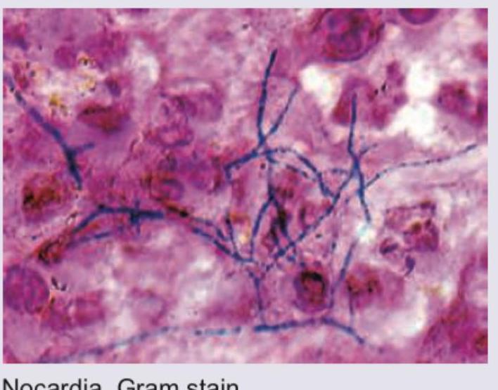
Stereotactic CT based aspiration from a patient with brain abscess was performed. Identify the organism seen and the stain used.

Explanation: **Explanation:** **Frei’s Test** is a delayed hypersensitivity skin test historically used for the diagnosis of **Lymphogranuloma Venereum (LGV)**, which is caused by *Chlamydia trachomatis* serotypes L1, L2, and L3. 1. **Why Chlamydia is correct:** The test involves the intradermal injection of an antigen (originally derived from the pus of a bubo or grown in a yolk sac). A positive result is indicated by an inflammatory nodule (≥7mm) appearing within 48–72 hours. While it was the gold standard for decades, it is now largely obsolete, replaced by more sensitive and specific methods like Nucleic Acid Amplification Tests (NAAT). 2. **Why other options are incorrect:** * **Mycoplasma:** Diagnosis usually relies on PCR or serology (Cold agglutinins). * **Rickettsia:** Diagnosed via the Weil-Felix reaction (heterophile agglutination) or immunofluorescence assays. * **Sarcoidosis:** The specific skin test for Sarcoidosis is the **Kveim-Siltzbach test**, which uses sarcoidal spleen tissue. **High-Yield Clinical Pearls for NEET-PG:** * **LGV Presentation:** Characterized by a painless primary lesion followed by painful inguinal lymphadenopathy (the "Groove sign"). * **Antigen Specificity:** The Frei test antigen is genus-specific (common to all *Chlamydia* species), not species-specific. * **Other "Named" Tests in Microbiology:** * **Dick Test:** Scarlet Fever (*S. pyogenes*) * **Schick Test:** Diphtheria (*C. diphtheriae*) * **Mitsuda Test:** Leprosy (*M. leprae*) * **Casoni’s Test:** Hydatid disease (*E. granulosus*)
Explanation: ### Explanation **Correct Option: C. Previous infection alters Widal test results.** The Widal test is a tube agglutination test that detects antibodies (anti-O and anti-H) against *Salmonella Typhi* and *Paratyphi*. In individuals who have had a previous infection, **anamnestic responses** occur. Memory B-cells can cause a rapid rise in antibody titers during any subsequent unrelated febrile illness, leading to a false-positive result. Therefore, a single positive Widal test is often unreliable for diagnosing an acute infection. **Analysis of Incorrect Options:** * **A. The Widal test is confirmative in endemic areas:** In endemic areas, a significant portion of the healthy population may carry baseline antibodies. Thus, a single test is never confirmative; only a **four-fold rise** in titers in paired sera (collected 7–10 days apart) is diagnostic. * **B. Antibiotic treatment does not alter Widal test results:** Early administration of antibiotics (like Chloramphenicol or Ciprofloxacin) can suppress the immune response, leading to low or absent antibody titers, potentially causing a false-negative result. * **D. The Widal test is not altered by prior vaccination:** Immunization with the TAB vaccine or older parenteral typhoid vaccines induces high levels of H and O antibodies, which can persist for months, interfering with the interpretation of the test. **High-Yield Clinical Pearls for NEET-PG:** * **Timing:** Widal becomes positive only after the **first week** of fever (maximal sensitivity in the 2nd–3rd week). * **Diagnostic Titers:** Generally, **O > 1:80** and **H > 1:160** are considered significant, but this varies by local endemicity. * **Gold Standard:** Bone marrow culture is the most sensitive; Blood culture is the investigation of choice in the 1st week (**BASU** mnemonic: **B**lood-1st week, **A**ntibody/Widal-2nd, **S**tool-3rd, **U**rine-4th).
Explanation: ### Explanation The diagnosis of Typhoid (Enteric) fever depends on the duration of the illness. This patient presents in the **second week** of symptoms, making the **Widal test** the most appropriate investigation for confirmation. #### 1. Why the Widal Test is Correct The Widal test is a serological test that detects antibodies (Anti-O and Anti-H) against *Salmonella typhi*. These antibodies typically appear in the blood at the end of the first week and reach diagnostic titers during the **second and third weeks** of the fever. A significant titer (usually >1:160 for O and >1:160 for H) or a four-fold rise in paired sera confirms the diagnosis. #### 2. Why Other Options are Incorrect * **Blood Culture (Option C):** This is the gold standard in the **first week** of illness (90% sensitivity). By the second week, the sensitivity drops significantly as the bacteremia subsides. * **Stool Culture (Option B):** This is most useful during the **second and third weeks**, but it is often used to identify carriers or for late-stage diagnosis. It is less definitive than serology for acute confirmation in the second week. * **Urine Culture (Option A):** This becomes positive only in the **third week** of the illness. #### 3. NEET-PG High-Yield Pearls: "BASU" Rule To remember the investigation of choice by week, use the mnemonic **BASU**: * **Week 1:** **B**lood Culture (Earliest positive) * **Week 2:** **A**ntibody test (Widal Test) * **Week 3:** **S**tool Culture * **Week 4:** **U**rine Culture * **Most sensitive overall:** Bone marrow culture (remains positive even after starting antibiotics). * **Specific Media:** Wilson and Blair’s Bismuth Sulfite Agar (jet black colonies). * **Carrier state:** Regarded as "Permanent" if shedding persists for >1 year; detected via **Vi agglutination test**.
Explanation: **Explanation:** The correct answer is **Cholera**. The fundamental principle for transfusion-transmitted infections (TTIs) is that the pathogen must have a **viremic, bacteremic, or parasitemic phase** in the host's blood to be collected during donation. **Why Cholera is the correct answer:** * **Cholera** is caused by *Vibrio cholerae*, which is an **enterotoxin-mediated** non-invasive infection. The bacteria remain confined to the intestinal lumen and do not enter the bloodstream. Transmission occurs exclusively via the **fecal-oral route** (contaminated food/water). Therefore, it cannot be transmitted through blood. **Why the other options are incorrect:** * **West Nile Virus (WNV):** This is a known TTI. It causes a period of asymptomatic viremia in humans, allowing it to be transmitted via blood products and organ transplants. * **Toxoplasmosis:** *Toxoplasma gondii* can be transmitted via blood transfusion (specifically through infected leukocytes) or organ transplantation, particularly in immunocompromised recipients. * **Hepatitis B Virus (HBV):** This is one of the most common TTIs globally. It is a highly infectious blood-borne pathogen routinely screened for in all blood banks using HBsAg and Nucleic Acid Testing (NAT). **NEET-PG High-Yield Pearls:** * **Mandatory Screening in India:** Blood banks must screen for five infections: HIV, HBV, HCV, Syphilis (Treponema pallidum), and Malaria. * **Bacterial Contamination:** Platelets are the most common blood component associated with bacterial sepsis because they are stored at room temperature (20-24°C). * **Emerging TTIs:** Keep an eye on Zika virus, Babesiosis, and Leishmaniasis as potential transfusion risks often discussed in recent literature.
Explanation: **Explanation:** The correct answer is **D**. Tetanus is caused by *Clostridium tetani*, an anaerobic, spore-forming bacterium. It is **not** transmitted via respiratory droplets. Instead, transmission occurs through the contamination of wounds (lacerations, punctures, or umbilical stumps) with soil, dust, or animal feces containing bacterial spores. Once in an anaerobic environment, the spores germinate and release tetanospasmin, leading to the clinical manifestation of "lockjaw." **Analysis of other options:** * **Option A (Legionella):** This is a true statement. *Legionella pneumophila* is typically transmitted by inhaling contaminated **aerosols** from water systems like cooling towers, humidifiers, or showerheads. It does not spread person-to-person. * **Option B (Listeria):** This is a true statement. *Listeria monocytogenes* is unique because it is a **psychrophile**, meaning it can grow at refrigeration temperatures (4°C). Common vehicles include deli meats and unpasteurized soft cheeses. * **Option C (Leptospirosis):** This is a true statement. It is a classic zoonosis transmitted through skin or mucosal contact with **water or soil contaminated by the urine** of infected reservoir hosts, most commonly rats. **NEET-PG High-Yield Pearls:** * **Tetanus:** Look for "drumstick appearance" on microscopy. The toxin (tetanospasmin) acts by blocking the release of inhibitory neurotransmitters **GABA and Glycine** from Renshaw cells. * **Listeria:** Exhibits **"tumbling motility"** at 25°C and is a common cause of neonatal meningitis. * **Leptospirosis:** Often presents with conjunctival suffusion; severe form with jaundice and renal failure is known as **Weil’s Disease**.
Explanation: ### Explanation The diagnosis of **Typhoid (Enteric) Fever**, caused by *Salmonella Typhi*, follows a specific chronological pattern of positivity across different diagnostic modalities. **Why Blood Culture is Correct:** During the **first week** of illness, the primary pathological event is **bacteremia** (bacteria entering the bloodstream after multiplying in the Peyer's patches and mesenteric lymph nodes). Therefore, blood culture is the gold standard and the most sensitive test (70-90% positivity) during this initial phase. **Analysis of Incorrect Options:** * **Serum Widal Test:** This is a serological test that detects antibodies (Anti-O and Anti-H). These antibodies only reach significant titers by the **end of the second week**. Testing in the first week often yields false negatives. * **Stool Culture:** While *S. Typhi* is excreted in feces, it is most consistently positive during the **second and third weeks** of illness due to the shedding of bacteria from the gallbladder into the intestine. * **Urine Culture:** This is typically positive only in the **third week** of illness, following prolonged bacteremia and renal seeding. **NEET-PG High-Yield Pearls:** * **Mnemonic "BASU":** To remember the sequence of positivity: **B**lood (1st week) > **A**ntibody/Widal (2nd week) > **S**tool (3rd week) > **U**rine (4th week). * **Bone Marrow Culture:** This is the **most sensitive** overall (up to 95%) and remains positive even if the patient has already started antibiotics. * **Castaneda’s Medium:** A biphasic medium often used for blood cultures to reduce the risk of contamination during subculturing. * **Rose Spots:** These clinical findings (faint pink macules on the trunk) typically appear in the second week.
Explanation: **Explanation:** The VDRL (Venereal Disease Research Laboratory) test is a non-specific screening test for Syphilis that detects **reagin antibodies** (IgM and IgG) directed against a cardiolipin-cholesterol-lecithin antigen. Because cardiolipin is a normal component of mitochondrial membranes, any condition causing significant tissue damage or immune dysregulation can lead to a **Biological False Positive (BFP)** result. **Why Pregnancy is the Correct Answer:** In the context of this specific question and standard NEET-PG patterns, **Pregnancy** is considered a physiological state rather than a pathological one. While some older texts listed pregnancy as a cause of BFP, modern evidence and standardized examinations categorize it as a "transient" cause (lasting <6 months). However, when compared against the other options which are classic, high-intensity pathological triggers for BFP, Pregnancy is the "least likely" or the "except" choice in many competitive formats. *Note: In some classifications, pregnancy is a known cause of transient BFP, but in the hierarchy of medical exams, the other three are definitive, high-yield pathological causes.* **Analysis of Incorrect Options:** * **Lepromatous Leprosy:** A classic cause of chronic BFP due to massive tissue destruction and polyclonal B-cell activation. * **Infectious Mononucleosis:** An acute viral infection (EBV) that causes transient BFP through non-specific immune stimulation. * **HIV:** Chronic immune activation and B-cell dysfunction in HIV patients frequently lead to false-positive non-treponemal tests. **Clinical Pearls for NEET-PG:** * **BFP Causes (Mnemonic: "PRACTISE"):** **P**regnancy (Transient), **R**at-bite fever, **A**utoimmune (SLE - most common chronic cause), **C**hancroid, **T**ypus/TB, **I**nfectious mononucleosis/IV drug use, **S**yphilis (True positive), **E**nteric fever. * **Confirmatory Test:** Any positive VDRL must be confirmed with a specific treponemal test like **FTA-ABS** or **TPHA**. * **Prozone Phenomenon:** Can cause a false *negative* VDRL in secondary syphilis due to very high antibody titers.
Explanation: ### Explanation **Correct Option: C. Jarisch-Herxheimer reaction** The Jarisch-Herxheimer reaction (JHR) is an acute inflammatory response that occurs shortly after starting antimicrobial therapy for spirochetal infections (most commonly **Syphilis**, but also Lyme disease and Leptospirosis). * **Mechanism:** When antibiotics (typically Penicillin) kill a large number of spirochetes, the **lysis of bacterial cell walls** leads to the sudden release of endotoxin-like products (lipoproteins) and necrotic cell debris into the bloodstream. This triggers a massive release of cytokines (TNF-α, IL-6, and IL-8), resulting in symptoms like fever, chills, rigors, hypotension, and headache. **Why Incorrect Options are Wrong:** * **A. Arthus reaction:** A localized **Type III Hypersensitivity** reaction involving the deposition of antigen-antibody complexes in small blood vessels, typically following a booster vaccination. It is not caused by bacterial lysis. * **B. Serum sickness:** A systemic **Type III Hypersensitivity** reaction occurring after exposure to foreign proteins (e.g., antivenom). It presents with fever, rash, and polyarthritis 1–2 weeks after exposure. * **D. Infectious mononucleosis-ampicillin reaction:** A characteristic maculopapular rash that occurs in patients with EBV infection (Glandular fever) when they are mistakenly treated with **Ampicillin or Amoxicillin**. It is an immune-mediated drug eruption, not a result of bacterial lysis. **NEET-PG High-Yield Pearls:** * **Timing:** JHR usually occurs within **2–24 hours** of the first dose of antibiotics. * **Management:** It is self-limiting. Treatment is symptomatic with **NSAIDs**. * **Prevention:** In high-risk cases (e.g., neurosyphilis), corticosteroids may be used to blunt the cytokine storm. * **Classic Scenario:** A patient treated for Primary or Secondary Syphilis develops sudden high fever and worsening of skin lesions a few hours after a Penicillin G injection.
Explanation: **Explanation:** **Brucellosis**, caused by the genus *Brucella*, is a zoonotic infection transmitted to humans through direct contact with infected animals or consumption of unpasteurized dairy products. **Brucella melitensis** (primarily found in goats and sheep) is the most common and virulent species causing human disease. The disease is known by several synonyms based on its clinical presentation and historical geographical prevalence: * **Malta Fever:** Named after the island where it was first identified. * **Undulant Fever:** Characterized by a rising and falling (wave-like) temperature pattern. * **Mediterranean Fever:** Reflecting its endemicity in the Mediterranean basin. **Analysis of Incorrect Options:** * **Treponema pallidum:** The causative agent of **Syphilis**, a sexually transmitted infection. * **Borrelia burgdorferi:** The causative agent of **Lyme disease**, transmitted by *Ixodes* ticks, typically presenting with Erythema migrans. * **Pseudomonas aeruginosa:** An opportunistic pathogen causing nosocomial infections (pneumonia, UTI, sepsis), particularly in cystic fibrosis and burn patients. **High-Yield Clinical Pearls for NEET-PG:** * **Diagnosis:** The **Standard Agglutination Test (SAT)** is the most common serological test (significant titer >1:160). * **Culture:** *Brucella* is fastidious; **Castaneda’s medium** (biphasic medium) is the traditional culture method of choice. * **Complications:** Osteoarticular involvement (sacroiliitis) is the most common complication. * **Treatment:** WHO recommends **Rifampicin + Doxycycline** for 6 weeks. For neurobrucellosis or endocarditis, triple therapy (including Streptomycin) is used.
Explanation: ***Correct: Nocardia, Gram stain*** - The image displays **Gram-positive, branching filamentous bacteria**, which are characteristic morphological features of *Nocardia* species. These organisms are known to cause brain abscesses, especially in immunocompromised individuals. - The stain used is clearly a **Gram stain**, as evidenced by the purple/blue coloring of the bacterial cells (Gram-positive) against a pink/red background (counterstain). - *Nocardia* species are **weakly acid-fast** and show characteristic **branching, beaded filaments** that can be visualized with Gram staining. *Incorrect: Cryptococcus, India ink* - *Cryptococcus* is a **yeast** and would appear as round or oval budding cells, often with a prominent capsule, rather than branching filaments. - An **India ink stain** highlights the polysaccharide capsule of *Cryptococcus* as a clear halo against a black background, which is not what is seen in the image. *Incorrect: Streptococcus, Gram stain* - *Streptococcus* species are **Gram-positive cocci** that typically arrange in chains. They would appear as spherical cells in linear arrangements, not as filamentous structures. - While *Streptococcus* species can cause brain abscesses, their morphology in a Gram stain would be distinctly different from the branching filaments shown. *Incorrect: Staphylococcus, Gram stain* - *Staphylococcus* species are **Gram-positive cocci** that typically arrange in grape-like clusters. They would appear as spherical cells in irregular clumps, not as filamentous structures. - Similar to *Streptococcus*, their characteristic coccal morphology on a Gram stain would not match the filamentous structures observed in the image.
Respiratory Tract Infections
Practice Questions
Urinary Tract Infections
Practice Questions
Gastrointestinal Infections
Practice Questions
Skin and Soft Tissue Infections
Practice Questions
Central Nervous System Infections
Practice Questions
Bone and Joint Infections
Practice Questions
Cardiovascular Infections
Practice Questions
Sexually Transmitted Infections
Practice Questions
Zoonotic Infections
Practice Questions
Bloodstream Infections and Sepsis
Practice Questions
Fever of Unknown Origin
Practice Questions
Infections in Immunocompromised Host
Practice Questions
Get full access to all questions, explanations, and performance tracking.
Start For Free