Which of the following is typically seen in cerebrospinal fluid (CSF) in tubercular meningitis?

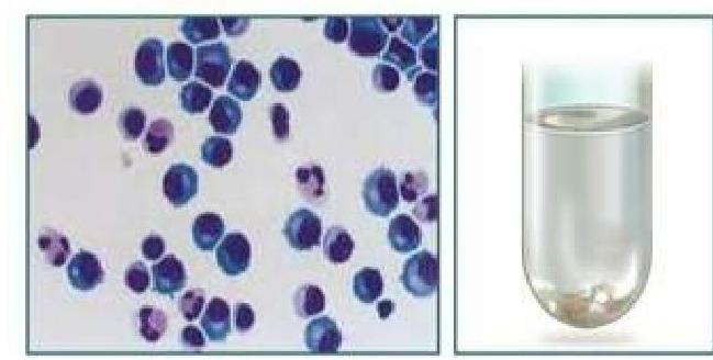
A patient comes to ED with fever and headache. On examination he has neck stiffness. CSF analysis was done: Glucose 55 mg/dl (normal 50-80), Protein 0.50 g/L (normal 0.18-0.45), ICT 35 cmH2O (normal 5-20), WBC 25 (predominantly lymphocytes). Most likely diagnosis is:
CSF in meningococcal meningitis shows -
Herpes virus involves which lobe?
A patient with a history of chronic ear infection now presents with manifestations, including headache and vomiting. A CT brain image is shown. What is the most probable diagnosis?

Brain abscesses in cyanotic heart disease are commonly located in
All of the following statements are true regarding central nervous system infections, except:
What is the most common cause of meningoencephalitis in children?
A young child with recurrent bacterial meningitis should be clinically evaluated for the presence of
Which of the following is the MOST accurate statement about CSF?
Explanation: ***Low opening pressure*** - While **elevated opening pressure** is common in tubercular meningitis due to inflammation and impaired CSF absorption, **low opening pressure** is not a typical finding. - Low opening pressure can occur with conditions like **CSF leak** or **spinal tap headache**, which are not directly characteristic of tubercular meningitis. *Low sugar* - **Low CSF glucose** (hypoglycorrhachia) is highly characteristic of bacterial and tubercular meningitis, as the bacteria or mycobacteria consume glucose. - The **CSF-to-blood glucose ratio** is typically less than 0.4 in tubercular meningitis. *High protein* - **Elevated CSF protein** is a consistent finding in tubercular meningitis, resulting from inflammation, increased vascular permeability, and breakdown of the blood-brain barrier. - Protein levels often range from **100-500 mg/dL** or even higher. *Lymphocytic Pleocytosis* - **Lymphocytic pleocytosis** (increased lymphocytes in CSF) is a hallmark of tubercular meningitis, although early stages might show a mixed cellular response. - The cell count typically ranges from **100-500 cells/µL**, predominantly lymphocytes.
Explanation: ***Coxsackie*** - The CSF analysis shows **normal glucose**, **mildly elevated protein**, **moderately elevated opening pressure**, and a **淋巴细胞主导** pleocytosis, which are characteristic findings in **viral meningitis**, commonly caused by enteroviruses such as Coxsackie virus [1]. - The combination of **fever, headache, neck stiffness**, and the specific CSF profile strongly points towards a viral etiology [1]. *Cryptococcus* - While fungal meningitis can present with similar symptoms and lymphocyte-dominant pleocytosis, it typically causes **markedly low CSF glucose** and **higher protein levels** than observed here. - Diagnosis usually requires specific tests like **India ink stain** or **cryptococcal antigen detection**, which are not indicated by these CSF findings. *TB* - Tuberculous meningitis usually presents with **very low CSF glucose** (often <40 mg/dl), **markedly elevated protein** (>1 g/L), and predominantly **lymphocytic pleocytosis**, often with a very slow onset [2]. - The CSF profile in this case, particularly the normal glucose, makes TB less likely [2]. *N. Gonorrhea* - *Neisseria gonorrhoeae* can cause **meningitis**, but it is generally a rare presentation and usually results in **neutrophilic pleocytosis** in the CSF, similar to other bacterial meningitides. - The **lymphocytic predominance** in this patient's CSF makes *N. gonorrhoeae* an unlikely cause.
Explanation: ***Gm '-ve' Diplococci in pus cells*** - Meningococcal meningitis is caused by *Neisseria meningitidis*, which are characteristic **Gram-negative diplococci**. - In active infection, these bacteria are typically found **intracellularly within polymorphonuclear leukocytes** (pus cells) in the cerebrospinal fluid. *Gm '+ve' bacilli* - This description would suggest a Gram-positive rod-shaped bacterium, such as *Listeria monocytogenes*, which is not the causative agent of meningococcal meningitis. - The morphology and Gram stain reaction are incorrect for *Neisseria meningitidis*. *Gm '+ve' Diplococci, in pus cells* - This describes organisms like *Streptococcus pneumoniae*, which are **Gram-positive diplococci** and a common cause of bacterial meningitis. - However, meningococcal meningitis is specifically caused by **Gram-negative** organisms. *Gm '-ve' bacilli* - This describes Gram-negative rod-shaped bacteria, such as *Haemophilus influenzae* or *Escherichia coli*. - While these can cause meningitis, *Neisseria meningitidis* is specifically a **diplococcus** (spherical in pairs), not a bacillus (rod-shaped).
Explanation: ***Temporal*** - Herpes virus (specifically **HSV-1**) often causes **encephalitis**, and the **temporal lobes** are the most common and severely affected regions [1]. - This predilection for the temporal lobes often leads to characteristic symptoms such as **memory deficits**, **aphasia**, and **behavioral changes** [1]. *Occipital* - While encephalitis can be diffuse, direct and primary involvement of the **occipital lobe** as the main site of herpes simplex encephalitis is **uncommon**. - Conditions like **posterior reversible encephalopathy syndrome (PRES)** or **cerebral amyloid angiopathy** are more typically associated with occipital involvement. *Parietal* - Although the **parietal lobe** can be affected in severe or widespread encephalitis, it is **not the primary or most frequent site** of herpes simplex encephalitis. - Damage to the parietal lobe would typically result in **sensory deficits**, **spatial disorientation**, or **neglect**. *Frontal* - The **frontal lobes** can be involved in herpes simplex encephalitis, often as part of a more extensive disease affecting the temporal lobes. - However, the **temporal lobes are more consistently and severely impacted**, accounting for the majority of the characteristic clinical presentation [1].
Explanation: ***Temporal lobe Abscess*** - The CT scan shows a **ring-enhancing lesion** with significant surrounding edema, which is characteristic of a **brain abscess**. - Given the history of a **chronic ear infection**, the temporal lobe is a common site for bacterial spread from the mastoid air cells or middle ear. *Meningitis* - Meningitis involves inflammation of the **meninges** and typically presents with diffuse changes on imaging, such as sulcal effacement or leptomeningeal enhancement, rather than a focal, encapsulated lesion. - While it can cause headache and vomiting, the CT image does not show findings typical of meningitis. *Extradural Abscess* - An extradural (or epidural) abscess is located **between the dura mater and the skull bone**. - It would typically appear as a collection outside the brain parenchyma, potentially causing mass effect but distinct from an intraparenchymal lesion seen in the image. *Cerebral Abscess* - The image does show a **cerebral abscess**, but this option is less specific than "Temporal lobe abscess." - The question asks for the **most probable diagnosis**, and combining the imaging findings with the patient's history of ear infection points to a specific location within the cerebrum.
Explanation: ***Parietal lobe*** - Brain abscesses in patients with **cyanotic heart disease** most commonly occur in the **parietal lobe**, followed by the frontal lobe. - This is due to the **hematogenous spread** of bacteria through the shunts, with a preference for areas with higher **blood flow** and terminal arteriolar distribution. *Cerebellar hemisphere* - While cerebellar abscesses can occur, they are **less common** than supratentorial abscesses in cyanotic heart disease. - They are more frequently seen in cases of direct spread from **ear infections**. *Thalamus* - Abscesses in the thalamus are generally **rare** and not a typical site for brain abscesses associated with cyanotic heart disease. - The thalamus's deep location makes it less susceptible to **hematogenous seeding** compared to cortical areas. *Temporal lobe* - Temporal lobe abscesses are common, but they are more frequently linked to **otitis media** or other local infections rather than the systemic bacteremia associated with cyanotic shunts. - While possible, it's **less primary** than the parietal or frontal lobes in this specific context.
Explanation: ***Cytomegalovirus is a common cause of bilateral temporal lobe hemorrhagic infarction.*** - **Cytomegalovirus (CMV)** typically causes **ventriculoencephalitis or periventricular necrosis** and microglial nodules in immunocompromised patients, not bilateral temporal lobe hemorrhagic infarction. - **Herpes simplex virus type 1 (HSV-1)** is the classic infectious cause of **bilateral temporal lobe hemorrhagic infarction (necrotizing encephalitis)**. *Prions infection causes spongiform encephalopathy* - **Prions** are misfolded proteins that cause transmissible spongiform encephalopathies (TSEs), such as Creutzfeldt-Jakob disease, characterized by **neuronal loss** and vacuolation (spongiform changes). - These diseases are invariably fatal and lead to rapid neurological deterioration. *JC virus is causative agent for progressive multifocal leucoencephalopathy* - The **JC virus** specifically targets and destroys **oligodendrocytes**, the myelin-producing cells of the central nervous system. - This leads to **demyelination** in multiple areas of the brain, causing the characteristic lesions seen in progressive multifocal leukoencephalopathy (PML). *Measles virus is the causative agent for subacute sclerosing pan encephalitis (SSPE).* - **Subacute sclerosing panencephalitis (SSPE)** is a rare, fatal, progressive neurodegenerative disease caused by a persistent and defective **measles virus infection** in the brain. - It occurs years after the initial measles infection, leading to cognitive decline, seizures, and motor dysfunction.
Explanation: ***Enterovirus*** - **Enteroviruses** (including coxsackievirus and echovirus) are the **most common cause** of viral meningoencephalitis and aseptic meningitis in children overall, accounting for the majority of cases. - They typically cause **mild, self-limiting illness** with good prognosis, particularly during **summer and fall months**. - Highly transmissible via fecal-oral route and respiratory droplets. *HSV* - **Herpes simplex virus (HSV)** is the **most common cause of severe sporadic encephalitis** requiring specific treatment, especially HSV-1 in older children and HSV-2 in neonates. - Causes **focal necrotizing encephalitis** with temporal lobe involvement, leading to severe neurological sequelae if untreated. - While less common in total number of cases than enteroviruses, it represents the most important **treatable** cause of severe encephalitis. *Arbovirus* - Arboviruses (such as **West Nile virus**, **Japanese encephalitis**, or **La Crosse virus**) cause meningoencephalitis with significant geographic and seasonal variation. - Transmitted by **mosquitoes** or **ticks**, making them less common overall than enteroviruses globally. *Poliovirus* - Wild-type **poliovirus** is now **eradicated in most countries** due to successful vaccination programs, making poliovirus-related meningoencephalitis extremely rare. - Historically caused **paralytic poliomyelitis** with anterior horn cell involvement, but is not a relevant cause in the current era.
Explanation: ***Spina bifida occulta with a dermal sinus tract*** - A **dermal sinus tract** provides a direct pathway for skin flora to access deeper structures, including the **meninges**, leading to recurrent bacterial meningitis - This condition arises from incomplete closure of the neural tube and is often associated with cutaneous stigmata such as a **dimple, tuft of hair, or hemangioma** in the lumbosacral region - Clinical examination should focus on the **midline back** for these telltale signs - This is the **most common anatomical cause** of recurrent bacterial meningitis in children *Hypoplastic left heart syndrome* - This is a congenital heart defect resulting in an underdeveloped left side of the heart, leading to cyanosis and heart failure - It is not directly associated with an increased risk of recurrent bacterial meningitis - This is a **cardiac anomaly**, not a CNS communication defect *Syringomyelia of the lower cervical cord* - Syringomyelia involves the formation of a fluid-filled cavity (syrinx) within the spinal cord, typically causing neurological deficits related to pain, temperature sensation, and motor weakness - While it is a neurological condition, it does **not disrupt the meningeal barrier** and therefore does not explain recurrent bacterial meningitis - This is an **intramedullary lesion** without external communication *Holoprosencephaly* - This is a severe condition where the forebrain fails to develop into two hemispheres, leading to various craniofacial anomalies and significant neurological impairment - It is a developmental brain abnormality and is **not a cause** of recurrent bacterial meningitis - While severe, it does not create a pathway for bacterial entry into the CNS
Explanation: ***Formed by the choroid plexus in the ventricles.*** * The **choroid plexus**, located in the ventricles of the brain, is primarily responsible for the production of **cerebrospinal fluid (CSF)**. * Specialized epithelial cells of the choroid plexus filter blood plasma to produce CSF, which then circulates through the central nervous system. *Normally contains no neutrophils* * Normal CSF should contain **virtually no neutrophils**; their presence typically indicates an inflammatory or infectious process, such as **bacterial meningitis**. * While normal CSF doesn't have neutrophils, this option isn't as broadly accurate as the choroid plexus statement because the presence of other cell types like lymphocytes in small numbers is normal. *pH is less than that of plasma* * The pH of CSF is typically **slightly lower than that of plasma** (around 7.31 compared to 7.40), but the statement "less than" is broad and the degree of difference can be variable and is a less defining characteristic than its formation site. * This slight difference in pH is important for regulating **respiration** through chemoreceptors, but it's not the most accurate or fundamental statement about CSF properties. *Removal of CSF during dural tap can cause a headache due to the change in pressure.* * A **post-dural puncture headache** (PDPH) is a well-known complication of a dural tap (lumbar puncture), caused by the leakage of CSF from the puncture site, leading to **intracranial hypotension**, not simply a change in pressure. * This decrease in CSF volume and pressure causes a traction on pain-sensitive structures within the cranium, resulting in a headache that is typically **worse when upright** and relieved by lying down.
Get full access to all questions, explanations, and performance tracking.
Start For Free