
Rheumatology and Immunology — MCQs

On this page
A 45-year-old patient presents with skin thickening, Raynaud's phenomenon, and digital ulcers. Which enzyme is the target of autoantibodies in this autoimmune disorder?
A 35-year-old female with heliotrope rash and Gottron's papules has a muscle biopsy showing inflammatory myopathy. What is the diagnosis?
Ankylosing spondylitis most commonly affects which region of the spine?
A 54-year-old woman presents with proximal muscle weakness and a skin rash over her knuckles. Laboratory tests show elevated muscle enzymes and a positive anti-Jo-1 antibody. What is the most likely diagnosis?
A 60-year-old woman with rheumatoid arthritis presents with severe pain and swelling in her right knee. Arthrocentesis reveals cloudy fluid with a high white blood cell count. What is the most likely diagnosis?
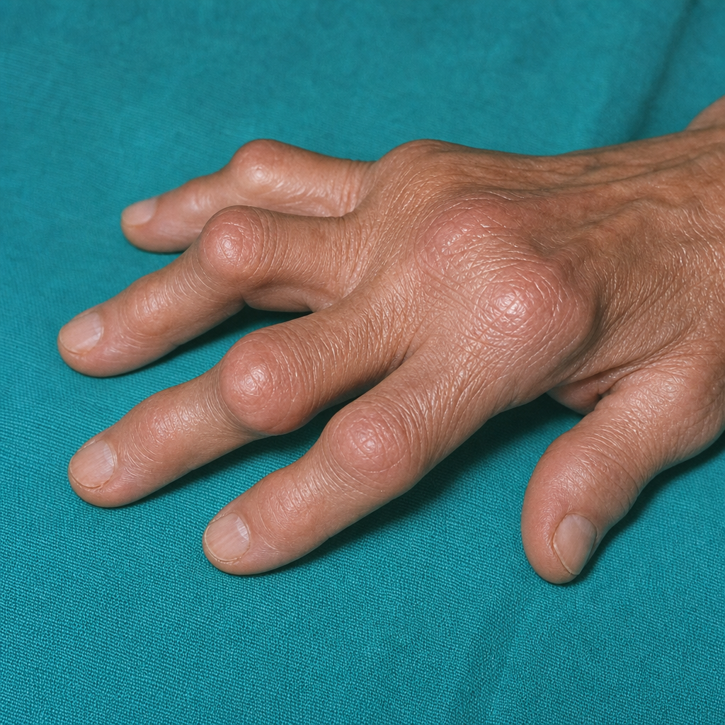
The patient is presenting with the deformity of the finger as shown. The PIP is involved but the DIP is spared
Which of the following is NOT a typical feature of seropositive rheumatoid arthritis?
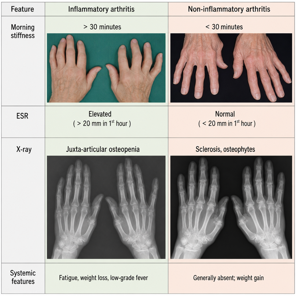
Which of the following is NOT a feature of inflammatory arthritis?
All are true about Marie-Strumpell disease except which of the following?
Psoriatic arthritis most commonly involves which joint -
Practice by Chapter
Rheumatoid Arthritis
Practice Questions
Spondyloarthropathies
Practice Questions
Systemic Lupus Erythematosus
Practice Questions
Vasculitis Syndromes
Practice Questions
Scleroderma and Related Disorders
Practice Questions
Inflammatory Myopathies
Practice Questions
Crystal Arthropathies
Practice Questions
Osteoarthritis
Practice Questions
Primary Immunodeficiency Disorders
Practice Questions
Autoinflammatory Syndromes
Practice Questions
Sjögren's Syndrome
Practice Questions
Antiphospholipid Syndrome
Practice Questions
Want unlimited practice?
Get full access to all questions, explanations, and performance tracking.
Scan to download app