
Rheumatology and Immunology — MCQs

On this page
"Telescope fingers" occur in
An elderly male presents with pain in his shoulders and hands. ESR is 105 mm/L. History includes transient blindness and unilateral headache.
Which of the following is a 'Small Vessel Vasculitis':
All of the following are found in SLE except
Antibody in drug induced Lupus
Which one of the following is not a diagnostic criterion for rheumatoid arthritis according to the American Rheumatism Association?
Sjogren's syndrome includes all except:
A 34-year-old woman has Raynaud's phenomenon associated with systemic sclerosis (scleroderma). Which of the following is the most appropriate management for this condition?
Which of the following manifestation of rheumatic fever disappears completely?
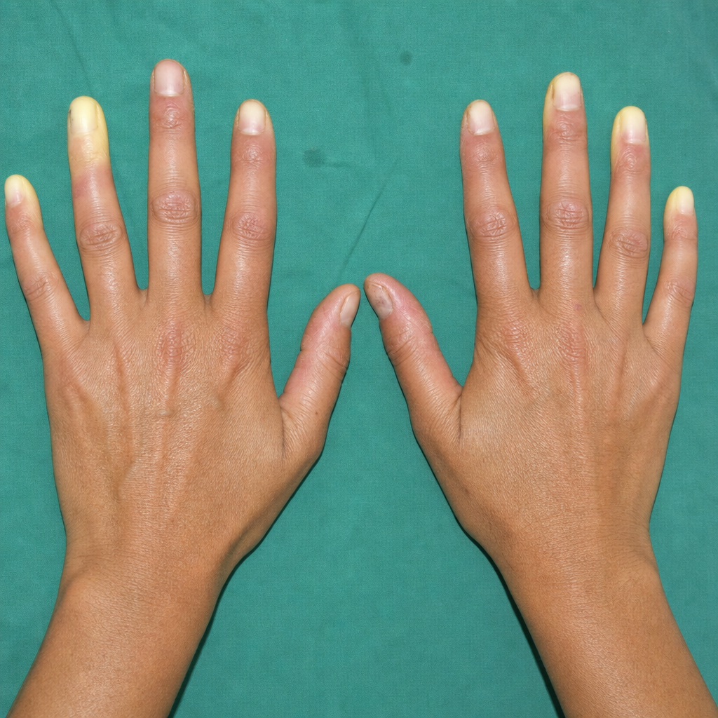
A 35 year old lady complains of dysphagia, Raynaud's phenomenon and her hands show the following appearance. Investigations show positive ANA. The likely diagnosis is
Practice by Chapter
Rheumatoid Arthritis
Practice Questions
Spondyloarthropathies
Practice Questions
Systemic Lupus Erythematosus
Practice Questions
Vasculitis Syndromes
Practice Questions
Scleroderma and Related Disorders
Practice Questions
Inflammatory Myopathies
Practice Questions
Crystal Arthropathies
Practice Questions
Osteoarthritis
Practice Questions
Primary Immunodeficiency Disorders
Practice Questions
Autoinflammatory Syndromes
Practice Questions
Sjögren's Syndrome
Practice Questions
Antiphospholipid Syndrome
Practice Questions
Want unlimited practice?
Get full access to all questions, explanations, and performance tracking.
Scan to download app