
Rheumatology and Immunology — MCQs

On this page
Characteristic feature of SLE is -
Arthritis mutilans is due to:
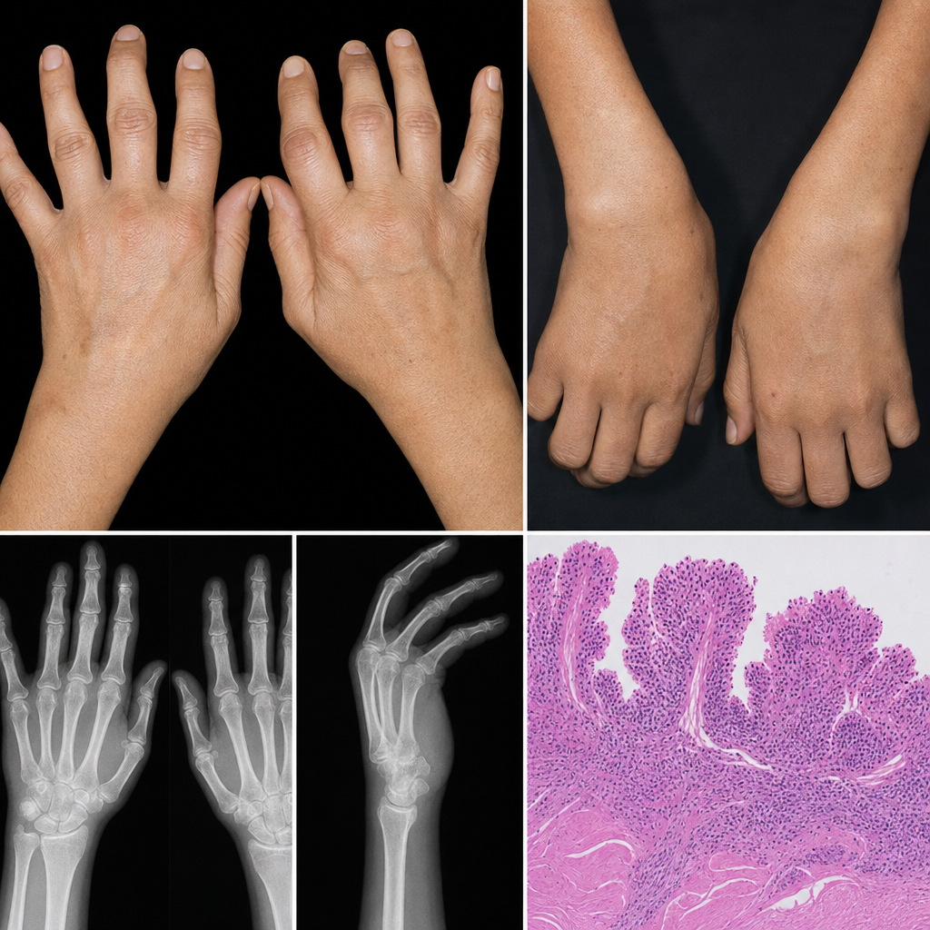
In rheumatoid arthritis, the characteristic joint involvement is:
Which of the following conditions results in arthritis without erosion of the bones?
An elderly male presents with pain in his shoulders and hips. Temporal arteries are tender to palpation. ESR is 105 mm/L.
What are the extra-articular manifestations of RA?
True statements about ankylosing spondylitis include all except: a) HLA-B27 is found in 90% of sufferers b) Uveitis is found in 15 to 20% of sufferers c) The condition is more common in females d) Radiological changes can occur in the spine before symptoms
A 40-year-old female presents with a history of fever, fatigue, weight loss, and polyarthralgia. Based on these symptoms, what is the most likely diagnosis?
About fibromyalgia all are true except
35 year old female patient complains of dry mouth and scratchy feeling in eyes. She is found to have antibodies against anti- SSA/Ro. Which of the following can be the most accurate sole criterion to diagnose the condition
Practice by Chapter
Rheumatoid Arthritis
Practice Questions
Spondyloarthropathies
Practice Questions
Systemic Lupus Erythematosus
Practice Questions
Vasculitis Syndromes
Practice Questions
Scleroderma and Related Disorders
Practice Questions
Inflammatory Myopathies
Practice Questions
Crystal Arthropathies
Practice Questions
Osteoarthritis
Practice Questions
Primary Immunodeficiency Disorders
Practice Questions
Autoinflammatory Syndromes
Practice Questions
Sjögren's Syndrome
Practice Questions
Antiphospholipid Syndrome
Practice Questions
Want unlimited practice?
Get full access to all questions, explanations, and performance tracking.
Scan to download app