
Rheumatology and Immunology — MCQs

On this page
What is the eye manifestation seen in HLA B27-positive patients with ulcerative colitis?
A 35-year-old patient presents with colicky abdominal pain, joint pain, and palpable purpura. Urinalysis shows only red blood cells with no other significant findings. Which of the following is the likely diagnosis?
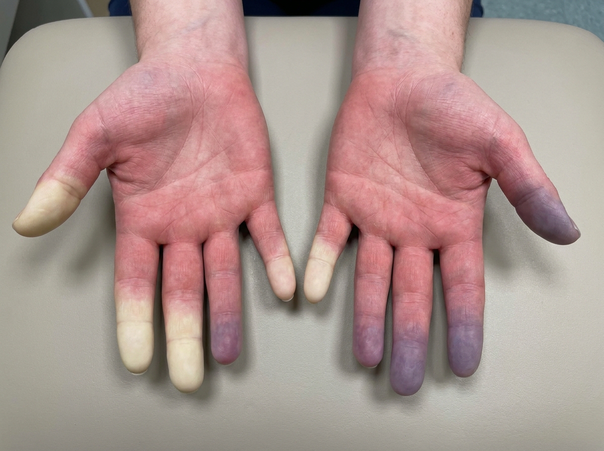
A 17-year-old woman with no comorbidities presents with numbness and paraesthesia of the fingers along with the characteristic finding as shown in the image below. She has no history of smoking or history of other illnesses. She mentions the episodes occur when she is under excess stress or during cold temperatures. What is the most likely diagnosis?
A 50-year-old woman presented with difficulty in activities like climbing stairs, getting up from a chair, and combing hair. Violaceous erythema of the upper eyelids was noted. What is the most probable diagnosis?
A 50-year-old female patient with a known case of ovarian cancer presents with difficulty in activities like climbing stairs, getting up from a chair, combing hair, etc. The following characteristic sign was found on examination. The most probable diagnosis is:
An elderly patient with the following deformity was brought to the OPD. What is the most probable diagnosis? 
A 35-year-old female presents with symmetrical joint pain and morning stiffness lasting 2 hours involving small joints of hands for 6 months. Examination reveals swelling of metacarpophalangeal and proximal interphalangeal joints bilaterally. ESR is 55 mm/hr, CRP is elevated, and rheumatoid factor is positive. X-ray shows periarticular osteopenia. What is the most appropriate initial treatment?
A patient with rheumatoid arthritis presents with neck pain and clinical signs of myelopathy. What is the most likely diagnosis?
A young man presents with chronic lower back pain and morning stiffness and pain on bending that improves with activity. X ray of LS spine is normal. Ocular examination shows anterior uveitis. Which diagnostic modality will pick up the disease process at the earliest?
A 20-year-old man presents with chronic back pain that is worse in the morning and improves with physical activity. He also has a history of anterior uveitis. A lumbar spine X-ray shows no abnormalities. Which of the following is the most appropriate next investigation for early diagnosis?
Practice by Chapter
Rheumatoid Arthritis
Practice Questions
Spondyloarthropathies
Practice Questions
Systemic Lupus Erythematosus
Practice Questions
Vasculitis Syndromes
Practice Questions
Scleroderma and Related Disorders
Practice Questions
Inflammatory Myopathies
Practice Questions
Crystal Arthropathies
Practice Questions
Osteoarthritis
Practice Questions
Primary Immunodeficiency Disorders
Practice Questions
Autoinflammatory Syndromes
Practice Questions
Sjögren's Syndrome
Practice Questions
Antiphospholipid Syndrome
Practice Questions
Want unlimited practice?
Get full access to all questions, explanations, and performance tracking.
Scan to download app