
Rheumatology and Immunology — MCQs

On this page
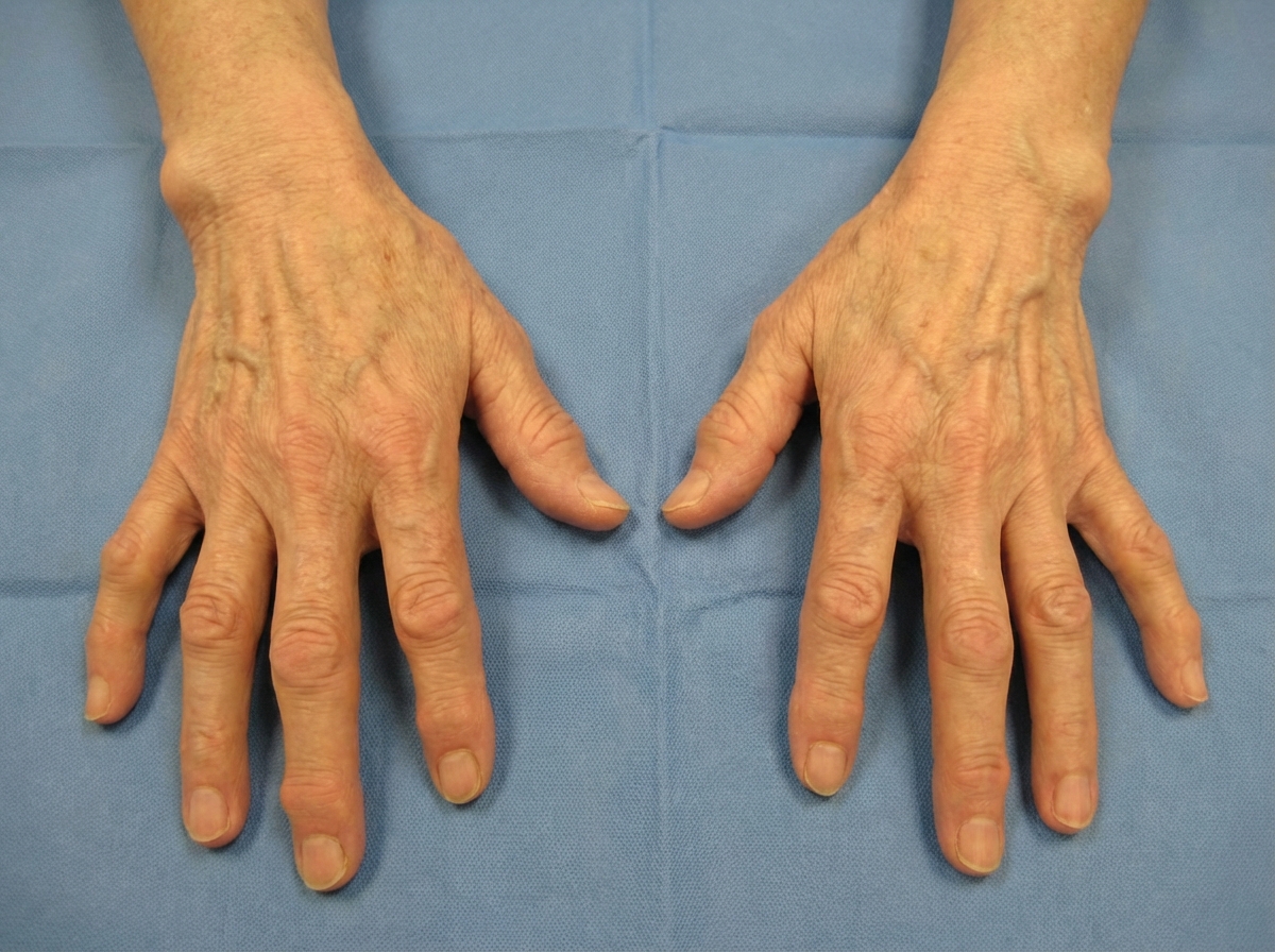
The deformity shown in the image is characteristic of which condition?
A patient presents with gritty pain in the eye and joint pain, following a recent urinary tract infection. What is the most probable diagnosis?
Which of the following conditions is associated with HLA-B27 in 90% of cases?
Which of the following extra-articular manifestations of Rheumatoid Arthritis is associated with an increased frequency of infections?
Sacro-iliac joint involvement is common in which condition?
A patient presented with arthritis and purpura. Laboratory examination showed monoclonal and polyclonal cryoglobulins. Histopathology showed deposits of cryoglobulins around the vessels. The patient should be tested for which of the following infections?
A 40-year-old man presents with a 2-week history of recurrent oral ulcers, genital ulcers, intermittent arthritic pain of the knees, and abdominal pain. Physical examination reveals shallow ulcerations of the mucosa of the glans penis, as well as oral aphthous ulcers and conjunctivitis. Which of the following is the most likely diagnosis?
A 45-year-old woman complains of severe headaches and difficulty in swallowing. Over the past 6 months, she has noticed small, red lesions around her mouth as well as thickening of her skin. The patient has "stone facies" on physical examination. Which of the following antigens is the most common and most specific target of autoantibody in patients with this disease?
Which of the following is NOT true about temporal arteritis?
Which HLA is associated with Reiter syndrome?
Practice by Chapter
Rheumatoid Arthritis
Practice Questions
Spondyloarthropathies
Practice Questions
Systemic Lupus Erythematosus
Practice Questions
Vasculitis Syndromes
Practice Questions
Scleroderma and Related Disorders
Practice Questions
Inflammatory Myopathies
Practice Questions
Crystal Arthropathies
Practice Questions
Osteoarthritis
Practice Questions
Primary Immunodeficiency Disorders
Practice Questions
Autoinflammatory Syndromes
Practice Questions
Sjögren's Syndrome
Practice Questions
Antiphospholipid Syndrome
Practice Questions
Want unlimited practice?
Get full access to all questions, explanations, and performance tracking.
Scan to download app