
Rheumatology and Immunology — MCQs

On this page
Which of the following medications is contraindicated in the treatment of an acute attack of gout?
A 30-year-old man presents with a 4-day history of cramping abdominal pain and bloody diarrhea. Physical examination reveals diffuse abdominal tenderness, normal bowel sounds, and no masses or organomegaly. Stool culture is positive for Shigella flexneri. The episode resolves spontaneously within one week. Six weeks later, he develops severe lower back pain, lumbar spine stiffness, and sacroiliac joint tenderness, treated with nonsteroidal anti-inflammatory agents. Two months after that, the back pain recurs, accompanied by right eye redness and blurred vision. Serologic testing for which of the following is most likely to be positive in this patient?
A 70-year-old retired military person with a good previous medical record complains of bi-temporal headache which is decreased in the lying down position. He states that he gets relief by applying pressure over the bilateral temples. The patient also complains of loss of appetite with a feverish feeling. What is the diagnosis?
Which of the following statements about polyarteritis nodosa (PAN) is FALSE?
Which of the following is NOT a feature of Ankylosing spondylitis (AS)?
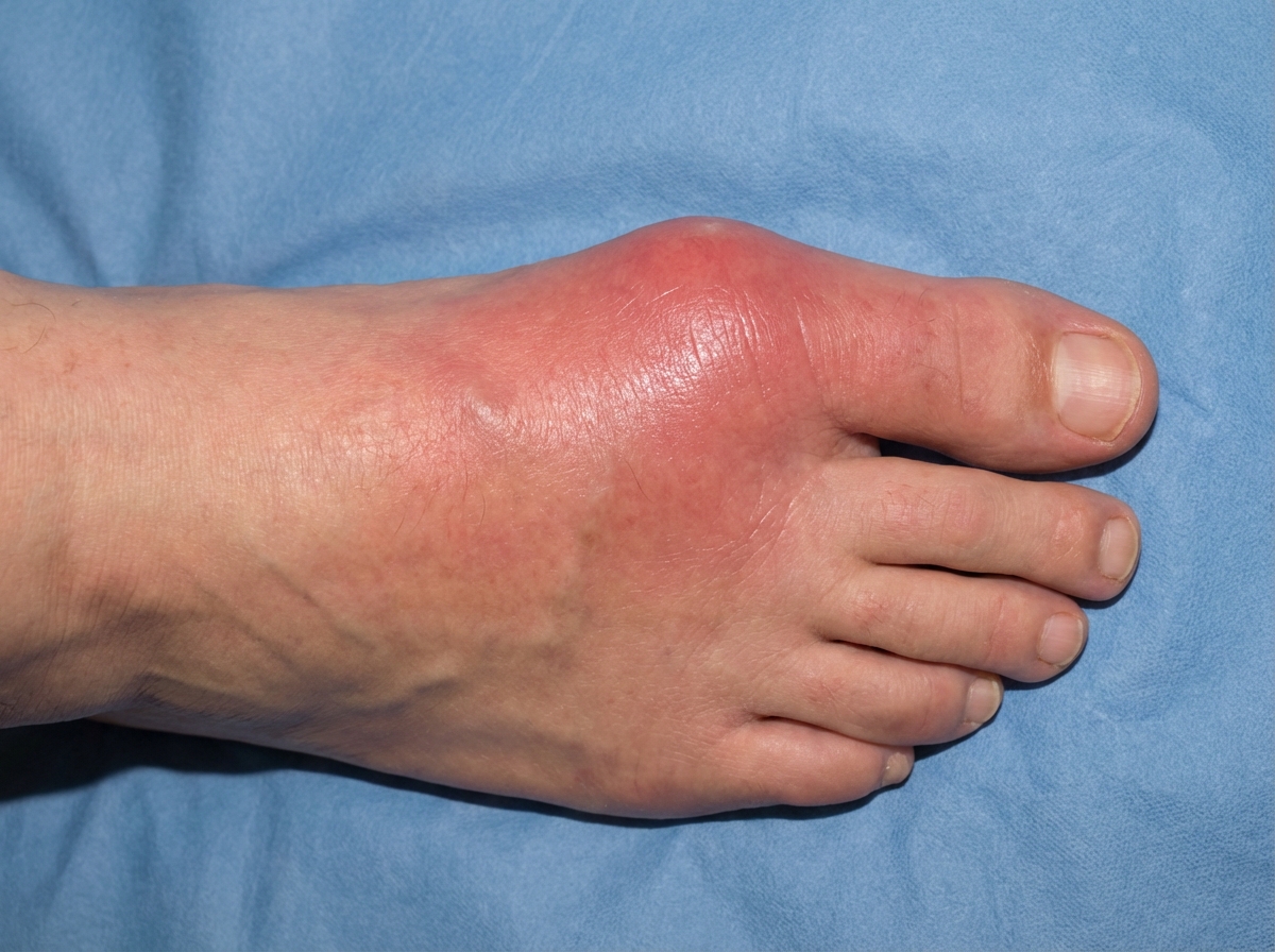
A 35-year-old man presents with acute onset of pain, swelling, and redness of his right big toe as shown in the image. What is the most likely diagnosis?
A patient presents with melaena, normal renal function, hypertension, and mononeuritis multiplex. What is the most probable diagnosis?
A patient with primary Sjogren syndrome treated with tear replacement for symptomatic relief notes continued parotid swelling for the last 3 months. She also has enlarged posterior cervical lymph nodes. Evaluation shows leukopenia and low C4 complement levels. What is the most likely diagnosis?
A 25-year-old male presented to the OPD with a complaint of recurrent oral ulcers and congested eyes. On enquiry, he has a history of prior hospital admission for venous thrombosis. What is the condition he is suffering from?
Which of the following is FALSE about Sjögren's syndrome?
Practice by Chapter
Rheumatoid Arthritis
Practice Questions
Spondyloarthropathies
Practice Questions
Systemic Lupus Erythematosus
Practice Questions
Vasculitis Syndromes
Practice Questions
Scleroderma and Related Disorders
Practice Questions
Inflammatory Myopathies
Practice Questions
Crystal Arthropathies
Practice Questions
Osteoarthritis
Practice Questions
Primary Immunodeficiency Disorders
Practice Questions
Autoinflammatory Syndromes
Practice Questions
Sjögren's Syndrome
Practice Questions
Antiphospholipid Syndrome
Practice Questions
Want unlimited practice?
Get full access to all questions, explanations, and performance tracking.
Scan to download app