
Rheumatology and Immunology — MCQs

On this page
A 67-year-old man with lung cancer presents with progressive weakness in his arms and legs. He reports difficulty rising from a chair and climbing stairs, with no associated pain or discomfort. Physical examination reveals proximal muscle strength of four out of five in both upper and lower limbs. His reflexes, muscle tone, and sensation are normal. He also has a heliotrope rash on his eyelids and Gottron's papules on his knuckles. Based on these findings, what is the most likely anatomic site of the disorder causing his muscle weakness?
A 50-year-old female presented with mild to moderate unilateral headache which becomes severe occasionally along with fever, jaw pain and visual disturbances, malaise, fatigue and weight loss. On examination, scalp tenderness was noted. Lab findings revealed normocytic normochromic anemia, elevated ESR and raised ANA titers. A biopsy was taken from the temporal artery. Which of the following cells are most importantly involved in the pathogenesis of the above disease?
Non-erosive arthritis is seen in which of the following conditions?
A 54-year-old man with a history of smoking presents with fever, hemoptysis, weight loss, and oligoarthritis. Serial skiagrams show fleeting opacities. What is the diagnosis?
In rheumatoid arthritis, which part of the spinal column is typically involved?
Which of the following is the most specific test for rheumatoid arthritis?
Circinate balanitis is a feature of which of the following conditions?
A 30-year-old male presents with numbness of both lower limbs and right upper limb. Examination reveals pulse 88/min and BP 160/110 mmHg. He also has digital gangrene involving the right 2nd and 3rd fingers. Urine routine examination is unremarkable. Microscopic examination shows RBCs. Hemogram and serum biochemistry are within normal limits. What is the most probable diagnosis?
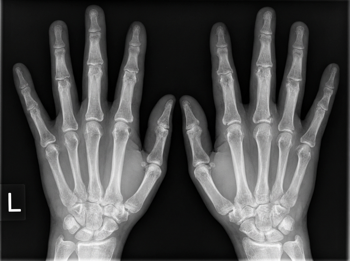
A 40-year-old female presents with a history of fever, weight loss, and polyarthralgia. Given the radiological findings, what is the most likely diagnosis?
Strawberry gingivitis is classically seen in which of the following conditions?
Practice by Chapter
Rheumatoid Arthritis
Practice Questions
Spondyloarthropathies
Practice Questions
Systemic Lupus Erythematosus
Practice Questions
Vasculitis Syndromes
Practice Questions
Scleroderma and Related Disorders
Practice Questions
Inflammatory Myopathies
Practice Questions
Crystal Arthropathies
Practice Questions
Osteoarthritis
Practice Questions
Primary Immunodeficiency Disorders
Practice Questions
Autoinflammatory Syndromes
Practice Questions
Sjögren's Syndrome
Practice Questions
Antiphospholipid Syndrome
Practice Questions
Want unlimited practice?
Get full access to all questions, explanations, and performance tracking.
Scan to download app