
Rheumatology and Immunology — MCQs

On this page
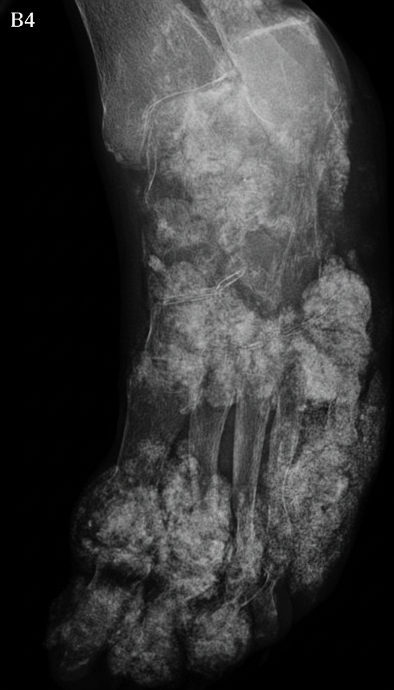
What is the most likely diagnosis of this case?
A 57-year-old man, previously asymptomatic and on no medications, develops a painful left big toe. It is so painful that even the weight of his sheets is excruciating. On examination, there is a swollen red toe. Which of the following medications is relatively contraindicated?
Which of the following is NOT true about ankylosing spondylitis?
Which of the following statements is FALSE regarding HIV-related arthritis?
In gout, tophi are typically found in which of the following locations?
Which one of the following disorders is not associated with carpal tunnel syndrome?
Which of the following conditions is characterized by non-deforming arthritis?
An 18-year-old boy presents with digital gangrene of the third and fourth fingers over the last 2 weeks. On examination, the blood pressure is 170/110 mm of Hg, and all peripheral pulses were palpable. Blood and urine examinations were unremarkable. ANA, Anti-dsDNA, and ANCA were negative. Which of the following represents the most likely diagnosis?
A 13-year-old female child presented with recurrent sinusitis, fever, arthralgia, respiratory distress, hematuria, and hypertension. Renal biopsy showed necrotizing granuloma. The anti-proteinase-3 ANCA was positive. What is the most likely diagnosis?
Which of the following statements is true about Rheumatoid arthritis?
Practice by Chapter
Rheumatoid Arthritis
Practice Questions
Spondyloarthropathies
Practice Questions
Systemic Lupus Erythematosus
Practice Questions
Vasculitis Syndromes
Practice Questions
Scleroderma and Related Disorders
Practice Questions
Inflammatory Myopathies
Practice Questions
Crystal Arthropathies
Practice Questions
Osteoarthritis
Practice Questions
Primary Immunodeficiency Disorders
Practice Questions
Autoinflammatory Syndromes
Practice Questions
Sjögren's Syndrome
Practice Questions
Antiphospholipid Syndrome
Practice Questions
Want unlimited practice?
Get full access to all questions, explanations, and performance tracking.
Scan to download app