
Rheumatology and Immunology — MCQs

On this page
A 40-year-old female presents with a history of fever, fatigue, weight loss, and symmetrical polyarthralgia predominantly involving the small joints of the hands and wrists. What is the most likely diagnosis?
Which of the following is seen more commonly in rheumatoid arthritis than in ankylosing spondylitis?
Bridge therapy is employed in which of the following conditions?
A 27-year-old male presents with burning micturition and urethral discharge. After 4 weeks, he develops joint pains involving both knees and ankles, redness of the eyes, and skin lesions. What is the most probable clinical diagnosis?
Which of the following statements is not true about Gout?
A 23-year-old man presents with new low back pain, stiffness, and left eye discomfort. He notes increased sensitivity to sunlight. The back pain is worse at night, described as a dull ache in the back and buttock area. Physical examination reveals paravertebral muscle, iliac crest, and ischial tuberosity tenderness with limited lumbar spine flexion. His eye is inflamed with a constricted pupil. Pelvic x-rays show sacroiliitis. What is the most likely diagnosis for his eye symptoms, considering it is the most common extra-articular manifestation of this condition?
A 27-year-old male presents with low backache that occurs early in the morning, is associated with stiffness, and persists for more than 30 minutes. On examination, his chest expansion is also restricted. What is the most probable diagnosis?
Which of the following statements is false regarding Rheumatoid Arthritis?
Which joint is NOT involved in Rheumatoid arthritis according to the 1987 ACR Classification Criteria?
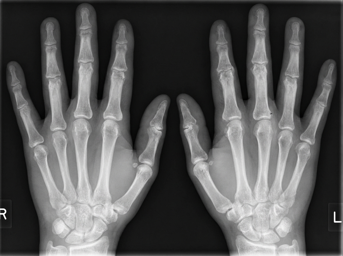
A 40-year-old female presents with a history of fever, weight loss, polyarthralgia, morning stiffness, and bilateral hand pain. Based on the provided radiological findings, what is the most likely diagnosis?
Practice by Chapter
Rheumatoid Arthritis
Practice Questions
Spondyloarthropathies
Practice Questions
Systemic Lupus Erythematosus
Practice Questions
Vasculitis Syndromes
Practice Questions
Scleroderma and Related Disorders
Practice Questions
Inflammatory Myopathies
Practice Questions
Crystal Arthropathies
Practice Questions
Osteoarthritis
Practice Questions
Primary Immunodeficiency Disorders
Practice Questions
Autoinflammatory Syndromes
Practice Questions
Sjögren's Syndrome
Practice Questions
Antiphospholipid Syndrome
Practice Questions
Want unlimited practice?
Get full access to all questions, explanations, and performance tracking.
Scan to download app