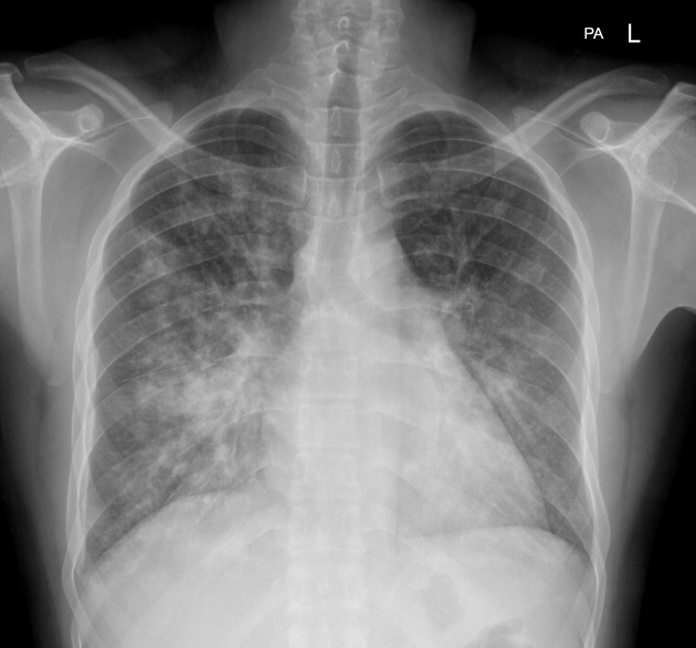
A 40-year-old man presented with repeated episodes of bronchospasm and hemoptysis. Chest X-ray revealed perihilar bronchiectasis. The most likely diagnosis is
Which of the following is NOT a characteristic feature of sarcoidosis?
Which of the following is NOT a contributing factor to pulmonary hypertension in COPD?
Pulmonary embolism is most commonly caused by:
Which statement is true regarding pneumothorax?
Which of the following statements about pulmonary embolism is false?
Hamman's sign is seen in:
Hemoptysis is said to be massive when it exceeds what amount in 24 hours?
Which of the following is the least likely cause of bronchiectasis?
Patient with pulmonary parenchymal fibrosis (PPF) who complains of breathing difficulty, is tachycardic, and tachypneic, and has a Batwing sign present on X-ray. What is the possible reason?

Explanation: ***Bronchopulmonary aspergillosis*** - **Allergic bronchopulmonary aspergillosis (ABPA)** is characterized by **bronchospasm**, recurrent fleeting pulmonary infiltrates, and **hemoptysis** in patients with asthma or cystic fibrosis. - The chest X-ray findings of **perihilar bronchiectasis** are a hallmark of ABPA, resulting from airway damage caused by the allergic response to *Aspergillus fumigatus*. *Sarcoidosis* - Sarcoidosis is a **granulomatous disease** that typically presents with non-caseating granulomas, often affecting the lungs, lymph nodes, eyes, and skin. [1] - While it can cause respiratory symptoms, **bronchiectasis** and **hemoptysis** are not typical primary features; more common findings include bilateral hilar lymphadenopathy and interstitial lung disease. [1] *Idiopathic pulmonary fibrosis* - Idiopathic pulmonary fibrosis (IPF) is a **chronic, progressive interstitial lung disease** characterized by progressive scarring of the lung tissue. [2] - While IPF causes respiratory symptoms like dyspnea and cough, it does not typically present with recurrent **bronchospasm**, **hemoptysis**, or **perihilar bronchiectasis**. [2] *Extrinsic allergic alveolitis* - Extrinsic allergic alveolitis, also known as **hypersensitivity pneumonitis**, is an immune-mediated lung disease caused by exposure to various inhaled antigens. - It typically presents with flu-like symptoms, cough, and dyspnea, and while it can cause interstitial lung disease, **bronchospasm** and **bronchiectasis** are not characteristic features.
Explanation: ***Erythema marginatum*** - This is a hallmark feature of **acute rheumatic fever**, characterized by a pink-red rash with raised borders and central clearing. - It is **not associated with sarcoidosis**, a multi-system granulomatous disease [1]. *Panda sign* - The **"Panda sign"** refers to increased uptake in the lacrimal and parotid glands on a gallium scan, a finding consistent with sarcoidosis. - This sign indicates **lymphocytic infiltration** of these glands, a common manifestation of the disease. *Egg shell calcification* - **Eggshell calcification** can be seen in the hilar or mediastinal lymph nodes in sarcoidosis, though it is more classically associated with **silicosis** [2]. - It represents calcification occurring in the periphery of a lymph node. *Hilar lymphadenopathy* - **Bilateral hilar lymphadenopathy** is the most common radiological finding in sarcoidosis, present in up to 90% of cases [1]. - It is often asymptomatic and detected on a routine chest X-ray [1].
Explanation: ***Bronchoconstriction*** - While common in COPD, **bronchoconstriction primarily increases airway resistance** and affects airflow, not directly pulmonary vascular pressure. - It does not directly cause the **remodeling** or **vasoconstriction** of pulmonary arteries that leads to sustained pulmonary hypertension. *Hypoxia* - **Chronic alveolar hypoxia** in COPD is a major driver of pulmonary hypertension by causing **pulmonary vasoconstriction**. - It also contributes to vascular remodeling, leading to sustained increases in pulmonary vascular resistance. *Pulmonary vasoconstriction* - **Hypoxia-induced pulmonary vasoconstriction** [1] is a primary and immediate response in the lungs that leads to increased pulmonary arterial pressure. - Over time, chronic vasoconstriction contributes to **vascular remodeling**, further exacerbating pulmonary hypertension. *High lung volume* - The **hyperinflation** characteristic of COPD can compress pulmonary capillaries and small vessels [2], leading to increased pulmonary vascular resistance. - This extrinsic compression contributes mechanically to the elevated pulmonary pressures seen in these patients.
Explanation: ***Deep vein thrombosis (DVT) of the leg*** - **Deep vein thrombosis (DVT)** in the leg is the most common source of emboli that travel to the lungs, leading to pulmonary embolism [1]. - The thrombus breaks off from the deep veins, typically in the **lower extremities**, and propagates through the venous system to the pulmonary arteries [1]. *Increased pulmonary pressure (a consequence of PE)* - **Increased pulmonary pressure** is a physiological consequence of a significant pulmonary embolism, as blood flow is obstructed, but it is not the cause of the embolism itself. - This option describes a **downstream effect**, rather than the origin of the embolus. *Fat embolism from pelvic fracture* - **Fat embolisms** can occur after long bone fractures (especially pelvic or femur fractures) and surgeries, but they are a less common cause of PE compared to DVT. - While they can lead to pulmonary symptoms, the mechanism involves **fat globules** entering the circulation, distinct from a thrombus. *Cardiac emboli from heart disease* - **Cardiac emboli** typically originate from the heart (e.g., from atrial fibrillation, mural thrombi after myocardial infarction, or valvular disease) and usually cause **systemic emboli** leading to strokes or limb ischemia. - While rare, paradoxal emboli can occur via a patent foramen ovale but are not the leading cause of "pulmonary" embolism.
Explanation: ***Absent breath sounds*** * **Complete absence of breath sounds** on the affected side is a hallmark sign of a pneumothorax, indicating collapsed lung tissue and air in the pleural space. * Air in the pleural space attenuates sound transmission, leading to a significant reduction or **absence of vesicular breath sounds** [2]. *Decreased percussion note* * A pneumothorax typically leads to a **hyperresonant percussion note** due to the presence of air in the pleural space. * A decreased or dull percussion note is usually associated with conditions like **pleural effusion** or consolidation [3]. *Always needs chest tube insertion* * **Small, stable pneumothoraces** can often be managed with observation and oxygen, especially if the patient is asymptomatic. * Chest tube insertion is reserved for **larger pneumothoraces**, symptomatic patients, or those with tension pneumothorax [1]. *Tracheal tug* * **Tracheal deviation**, not tracheal tug, can be a sign of a **tension pneumothorax**, where the trachea is pushed away from the affected side [1]. * Tracheal tug, or upward movement of the trachea with inspiration, is more indicative of **severe airway obstruction** or respiratory distress.
Explanation: Most commonly presents within 2 weeks - This statement is **false** because while pulmonary embolism can occur at any time, its onset is not restricted to or "most commonly presents" within a two-week period. - The timing of presentation can vary widely depending on the predisposing factors and the specific event leading to the embolus formation and migration. *Dyspnea is the most common symptom* - **Dyspnea** (shortness of breath) is indeed the **most frequent symptom** reported by patients experiencing pulmonary embolism. - This symptom often occurs acutely and can range from mild to severe, correlating with the size and location of the embolism. *Pulmonary embolism often arises from deep vein thrombosis in the legs.* - The vast majority of **pulmonary emboli (PEs)** originate from **deep vein thromboses (DVTs)**, particularly those located in the large veins of the legs and pelvis [1, 2]. - These clots can detach and travel through the venous system to the heart and then into the pulmonary arteries [2]. *Longer survival time generally indicates a better chance of recovery.* - Patients who survive longer after a pulmonary embolism are more likely to have received timely treatment and may have experienced a less severe embolic event or have better underlying health. - Prolonged survival after the initial acute phase suggests the patient has overcome the immediate life-threatening aspects and is on the path to recovery, potentially with less long-term sequelae.
Explanation: ***Pneumomediastinum*** - **Hamman's sign** is a classic auscultatory finding in pneumomediastinum, characterized by **crunching, bubbling, or clicking sounds synchronized with the heartbeat**. - These sounds are heard due to the presence of **air in the mediastinum** being displaced by the contracting heart. *Pericardial effusion* - **Pericardial effusion** involves fluid accumulation around the heart, which typically causes muffled heart sounds and, in severe cases, signs of **cardiac tamponade**. - It does not produce the characteristic **crunching sound** associated with Hamman's sign. *Superior vena cava obstruction* - **Superior vena cava obstruction** is characterized by symptoms like **facial swelling**, **distended neck veins**, and dyspnea due to impaired venous return. - It does not lead to the presence of air in the mediastinum or the specific auscultatory findings of Hamman's sign. *Thyrotoxicosis* - **Thyrotoxicosis** is a hypermetabolic state with symptoms such as **tachycardia**, palpitations, and heat intolerance. - While it affects the cardiovascular system, it does not involve the presence of air in the mediastinum or the development of Hamman's sign.
Explanation: 600mL - Hemoptysis is considered **massive** when the volume of blood expectorated exceeds **600 mL within a 24-hour period**, or **100 mL per hour**, indicating a life-threatening emergency. - This threshold is crucial because it significantly increases the risk of **airway obstruction** and **hypovolemic shock**, which requires immediate medical intervention. *400mL* - While 400 mL in 24 hours is a significant amount of bleeding, it is generally not classified as **massive hemoptysis** according to most clinical definitions. - Significant hemoptysis can still be dangerous and require investigation, but it does not meet the criteria for immediately life-threatening massive hemoptysis. *500mL* - This volume is close to the threshold for massive hemoptysis but is still typically not the **definitive cutoff** used in clinical practice. - It would still warrant aggressive management and consideration for interventions like bronchoscopy or embolization. *800mL* - While certainly a severe amount, 800 mL in 24 hours **exceeds** the generally accepted definition of massive hemoptysis. - The 600 mL threshold is based on the point at which there is a critical risk of **asphyxiation** or **hemodynamic instability** [1].
Explanation: Primary TB - While post-primary (reactivation) tuberculosis can lead to **bronchiectasis**, primary tuberculosis is less commonly a direct cause of established, widespread bronchiectasis in the acute phase. - Primary TB more often causes **lymphadenopathy** and localized parenchymal disease, with bronchiectasis being a rarer, late complication if airway obstruction or severe destruction occurs. *Pneumonia* - Severe or recurrent bacterial pneumonias can cause **irreversible damage** to the bronchial walls, leading to dilation and fibrosis, which is characteristic of bronchiectasis [1]. - The inflammatory process and **mucociliary dysfunction** during pneumonia contribute to ongoing infection and structural changes [1]. *Inhaled foreign body* - A persistent **foreign body** in the airway can cause localized obstruction, leading to chronic inflammation, infection, and subsequent dilation of the bronchi distal to the obstruction [1]. - This recurrent infection and inflammation weaken the bronchial walls over time, defining localized bronchiectasis. *Chronic aspiration* - **Recurrent aspiration** of gastric contents or oral secretions causes repeated injury and inflammation to the bronchial epithelium. - This chronic irritation and potential for bacterial colonization damage the airway walls, predisposing to **bronchial dilation** and infection, characteristic of bronchiectasis.
Explanation: ***Due to fluid overload in a patient with pulmonary parenchymal fibrosis*** - The presence of the **Batwing sign** (or butterfly pattern) on X-ray is characteristic of **pulmonary edema**, which is often caused by fluid overload. - In a patient with pre-existing **pulmonary parenchymal fibrosis (PPF)**, even moderate fluid overload can rapidly exacerbate respiratory symptoms and lead to acute pulmonary decompensation due to reduced lung compliance and impaired gas exchange. *Transfusion-related lung injury* - **Transfusion-related acute lung injury (TRALI)** typically presents with acute hypoxemia and bilateral infiltrates, which could resemble pulmonary edema, but often occurs within 6 hours of transfusion and is not directly linked to pre-existing pulmonary fibrosis in this context. - While TRALI can cause pulmonary edema, the scenario provided gives no information about recent transfusions, making fluid overload a more direct and common cause given the X-ray findings. *Acute renal failure due to tubular acidosis* - **Renal tubular acidosis** primarily affects acid-base balance and electrolyte levels, usually not directly causing acute, severe pulmonary symptoms or the "Batwing sign" unless there's associated severe fluid retention due to overall renal failure. - While acute renal failure can lead to fluid overload, tubular acidosis specifically points to a primary metabolic derangement rather than direct pulmonary involvement or the characteristic X-ray finding. *Hemolysis leading to hemoglobinuria* - **Hemolysis** can cause anemia and, in severe cases, acute kidney injury due to hemoglobinuria, but it does not directly explain acute respiratory distress, tachycardia, tachypnea, or a "Batwing sign" on chest X-ray. - These pulmonary findings are indicative of **fluid accumulation in the lungs**, which is not a direct consequence of hemolysis itself.
Obstructive Airway Diseases (Asthma, COPD)
Practice Questions
Interstitial Lung Diseases
Practice Questions
Pulmonary Infections
Practice Questions
Pulmonary Vascular Diseases
Practice Questions
Pleural Diseases
Practice Questions
Sleep-Disordered Breathing
Practice Questions
Respiratory Failure
Practice Questions
Mediastinal Disorders
Practice Questions
Occupational Lung Diseases
Practice Questions
Pulmonary Function Testing
Practice Questions
Bronchiectasis and Cystic Fibrosis
Practice Questions
Lung Cancer Approach
Practice Questions
Get full access to all questions, explanations, and performance tracking.
Start For Free