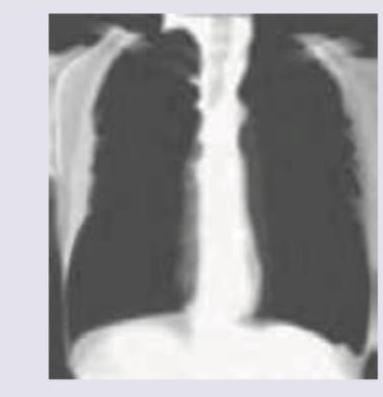
A 35-year-old coal worker presents with difficulty in breathing on exertion for last 2 years. CXR was performed. It shows:

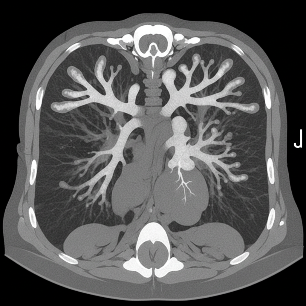
An asthmatic presents with brownish plugs in sputum. CT chest was performed. What is the diagnosis?

A 62-year old male chronic smoker has been diagnosed with Chronic Obstructive Pulmonary Disease (COPD). On pulmonary function testing, the ratio of Forced Expiratory Volume in 1 second (FEV1) to Forced Vital Capacity (FVC) was 0.6 and FEV1 was 70 % of predicted. What is the severity of airflow obstruction in this patient as per GOLD criteria?
Under the Stepwise Approach to the management of Bronchial Asthma, which one of the following is the correct initial treatment at Step 1 for a patient diagnosed with Asthma?
Consider the following statements: The clinical features of tension pneumothorax include 1. tracheal shift to contralateral side 2. absent breath sounds on the affected side 3. low output circulatory failure 4. peripheral cyanosis Which of the statements given above is/are correct?
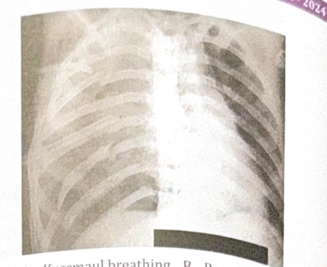
A 45-year-old male presents to the emergency department following a motor vehicle accident. He complains of severe chest pain and difficulty breathing. On examination, he appears distressed, with a respiratory rate of 28 breaths per minute. His oxygen saturation is 88% on room air. There is visible bruising on the chest, X-ray is done which is shown below. Which of the following is seen in this patient?

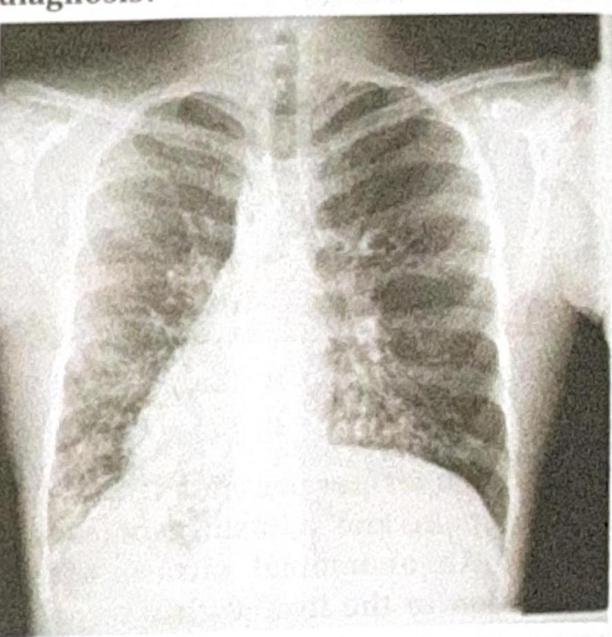
A 25-year-old patient presents with acute epigastric pain and elevated serum lipase. The patient was stabilized after 3 days, and a chest X-ray was obtained. What is the most common pulmonary complication associated with this condition?
A patient presents with respiratory distress and is diagnosed with panacinar emphysema. Which of the following is deficient?
A patient presents with recurrent lung infections, and the chest X-ray provided shows a characteristic finding. What is the most likely diagnosis?

Match the following: A) Caplan syndrome- 1) Found first in coal worker B) Asbestosis- 2) Upper lobe predominance C) Mesothelioma- 3) Involves lower lobe D) Sarcoidosis- 4) Pleural effusion is seen
Explanation: ***Reticulonodular infiltrates*** - The chest X-ray of a coal worker presenting with shortness of breath over two years is highly suggestive of **coal workers' pneumoconiosis (CWP)**. - CWP manifests as **reticulonodular infiltrates** on chest X-ray due to the accumulation of coal dust and the body's inflammatory response. *Emphysema* - **Emphysema** typically presents with **hyperinflated lungs** and **flattened diaphragms** on CXR, which are not clearly visible here. - While smoking can coexist with CWP and lead to emphysema, the primary CXR finding for CWP is interstitial changes. *Pulmonary fibrosis* - Although **pulmonary fibrosis** is characterized by reticular opacities, it often progresses to **honeycombing** and **volume loss**, which are not consistently or predominantly seen in earlier stages of CWP. - While CWP can lead to progressive massive fibrosis (PMF) in advanced stages, the initial description of reticulonodular infiltrates is more characteristic of simple CWP. *Cardiac atrophy* - **Cardiac atrophy** refers to the **wasting away of heart muscle** and would not be directly visible or implied by typical chest X-ray findings for a lung disease. - The presented image focuses on lung pathology, and there is no indication of cardiac atrophy.
Explanation: ***Allergic Bronchopulmonary Aspergillosis (ABPA)*** - The CT chest demonstrates **central bronchiectasis with high-attenuation mucoid impaction** producing the classic **"finger-in-glove" sign** — pathognomonic for ABPA - The clinical triad of **asthma + brownish mucus plugs (containing fungal hyphae of *Aspergillus fumigatus*) + central bronchiectasis on CT** is diagnostic - Additional CT features include **bronchial wall thickening**, mosaic attenuation, and air trapping peripherally - ABPA results from a **Type I and Type III hypersensitivity reaction** to *Aspergillus fumigatus* colonizing the airways of asthmatics and cystic fibrosis patients *Aspergilloma* - Aspergilloma (mycetoma) presents as a **fungal ball within a pre-existing pulmonary cavity** with the "Monod sign" (air crescent sign) on CT - It typically occurs in patients with **structural lung disease** (old TB cavities, bronchiectasis) and does NOT cause central bronchiectasis with mucoid impaction - Clinical: hemoptysis is the hallmark, NOT mucus plugs in an asthmatic *Pulmonary Tuberculosis* - TB on CT shows **upper lobe consolidation, tree-in-bud nodularity, cavitation**, and lymphadenopathy - Does NOT produce central bronchiectasis with mucoid impaction or the finger-in-glove pattern - Sputum would show **AFB**, not brownish mucus plugs from fungal colonization *Eosinophilic Pneumonia* - CT shows **bilateral peripheral consolidation** (reverse pulmonary edema pattern / "photographic negative of pulmonary edema") - Associated with peripheral blood eosinophilia and BAL eosinophilia - Does not produce mucoid impaction or central bronchiectasis; lacks the *Aspergillus* colonization component
Explanation: **Stage II - Moderate** - According to GOLD criteria, an FEV1/FVC ratio of less than 0.70 confirms airflow obstruction [1]. In this case, the ratio is 0.6. - A predicted FEV1 between 50% and 79% (inclusive) indicates **moderate COPD**, which aligns with the patient's FEV1 of 70% predicted [1]. *Stage III - Severe* - This stage is characterized by a **post-bronchodilator FEV1** between 30% and 49% of predicted [1]. - The patient's FEV1 of 70% predicted is too high for Stage III, indicating less severe obstruction. *Stage IV - Very severe* - This is the most severe stage, defined by a **post-bronchodilator FEV1** less than 30% of predicted, or FEV1 less than 50% predicted with signs of respiratory failure [1]. - The patient's FEV1 of 70% predicted is significantly higher than the threshold for very severe COPD. *Stage I - Mild* - Stage I is diagnosed when the **post-bronchodilator FEV1** is 80% or greater than predicted [1]. - The patient's FEV1 of 70% predicted falls below this criterion, indicating a more significant obstruction than mild.
Explanation: ***Low dose inhaled corticosteroid only*** - For newly diagnosed asthma patients requiring daily controller therapy (Step 1 or 2 as per GINA 2021+), a **low-dose inhaled corticosteroid (ICS)** is the recommended initial monotherapy [1]. - ICS addresses the underlying inflammation in asthma, which is crucial even in mild persistent cases. *Low dose inhaled corticosteroid plus leukotriene antagonist* - This combination is typically considered at **higher steps** (e.g., Step 3 or 4) if control is not achieved with low-dose ICS alone or if there are specific indications like **aspirin-exacerbated respiratory disease** [1]. - Initiating with two controller medications at Step 1 is generally not recommended as per guideline. *Low dose inhaled corticosteroid plus long acting anti-muscarinic agents* - **Long-acting muscarinic antagonists (LAMAs)** are primarily used in **severe asthma** that remains uncontrolled despite ICS/LABA therapy, usually at Step 4 or 5. - They are not considered a first-line addition to ICS at Step 1. *Low dose inhaled corticosteroid plus oral corticosteroid* - **Oral corticosteroids** are reserved for asthma **exacerbations** or very severe, uncontrolled asthma, used for short periods due to significant systemic side effects [1]. - They are never used as initial daily maintenance therapy at Step 1 due to their high side effect profile.
Explanation: ***1, 2 and 3*** - **Tracheal deviation to the contralateral side** is a hallmark sign of tension pneumothorax [1], caused by the increasing pressure in the affected hemithorax pushing mediastinal structures away. - **Absent breath sounds on the affected side** result from the complete collapse of the lung and inability of air to enter the pleural space with inspiration [1]. **Low output circulatory failure** occurs due to increased intrathoracic pressure compressing the heart and great vessels, impairing venous return and cardiac output [2]. *1 and 3 only* - While **tracheal shift to the contralateral side** and **low output circulatory failure** [2] are indeed features, this option incorrectly excludes **absent breath sounds on the affected side**, which is a critical clinical finding in tension pneumothorax. - The absence of breath sounds directly reflects the collapsed lung, a primary mechanical consequence of the condition. *2 and 4* - **Absent breath sounds on the affected side** is correct, but **peripheral cyanosis** is not a *primary* or *early* distinguishing feature of tension pneumothorax. While hypoxemia can lead to cyanosis, it's often a late and less specific sign. - This option misses the crucial finding of **tracheal deviation** and the systemic impact of **circulatory failure** [2], which are more direct indicators of the severity and mechanism of tension pneumothorax. *1 and 4* - **Tracheal shift to the contralateral side** is a correct feature. However, **peripheral cyanosis** is a less specific and often later sign compared to the direct mechanical and circulatory effects. - This option incorrectly omits **absent breath sounds on the affected side** and **low output circulatory failure**, both of which are more consistently present and diagnostically important in tension pneumothorax.
Explanation: ***Bronchial breathing*** - The provided image is a chest X-ray. The question describes a patient with severe chest pain, difficulty breathing, hypoxemia, and visible chest bruising after a motor vehicle accident, which suggests **pulmonary contusion** or **pneumonia**. - In such conditions, **bronchial breathing** might be heard on auscultation due to consolidation or compression of lung tissue. The X-ray shows diffuse infiltrates or opacities indicative of lung injury, making bronchial breathing a plausible physical exam finding. *Kussmaul breathing* - This is a pattern of **deep and labored breathing** associated with severe **metabolic acidosis**, particularly diabetic ketoacidosis. - While the patient is in distress, the chest X-ray and accident history point to acute respiratory injury rather than metabolic acidosis as the primary cause. *Paradisical breathing* - This term is **not a recognized medical breathing pattern**. It appears to be a distracter. - Medical terminology for breathing patterns includes terms like Kussmaul, Cheyne-Stokes, Biot's, etc. *Apneustic breathing* - Characterized by **prolonged inspiratory pauses** followed by short exhalations, indicating severe damage to the **pons (brainstem)**. - This breathing pattern is typically seen with neurological insults like stroke or severe head injury, which is not directly indicated by the information given, although a head injury could theoretically occur in a MVC, it's not the primary finding for the presented complaints and X-ray.
Explanation: ***ARDS (Acute Respiratory Distress Syndrome)*** - **Acute pancreatitis** can lead to significant systemic inflammation, causing widespread lung injury and subsequent **ARDS** [1, 2]. - This complication presents with bilateral infiltrates on chest X-ray [3] and severe **hypoxemia** refractory to oxygen therapy [1]. *Acute pulmonary edema* - While pulmonary edema can occur in critical illness, **cardiogenic pulmonary edema** is less common as the primary pulmonary complication of pancreatitis in young, otherwise healthy patients [3]. - Pancreatitis-related pulmonary issues are more often inflammatory, leading to lung injury rather than direct fluid overload from cardiac dysfunction. *Pulmonary thromboembolism* - Although critically ill patients, including those with acute pancreatitis, are at increased risk for **thrombotic events**, it's not the most common immediate pulmonary complication. - A pulmonary embolism would typically present with pleuritic chest pain, dyspnea, and potentially hemodynamic instability, and often without diffuse infiltrates on chest X-ray unless severe. *Miliary TB* - **Miliary tuberculosis** is a chronic infectious process characterized by widespread dissemination of Mycobacterium tuberculosis. - It would not manifest as an acute complication of acute pancreatitis and typically presents with constitutional symptoms and a different pattern on chest X-ray.
Explanation: **Alpha-1 antitrypsin** * **Alpha-1 antitrypsin (A1AT) deficiency** is a genetic disorder that leads to the development of panacinar emphysema, especially in non-smokers or at a young age [1], [2]. * A1AT protects the lung tissue from destruction by **elastase** released by neutrophils; without it, this enzyme breaks down alveolar walls [1], [2]. *Surfactant* * **Surfactant** is responsible for reducing surface tension in the alveoli, preventing their collapse in the lungs. * A deficiency primarily causes **neonatal respiratory distress syndrome** or adult respiratory distress syndrome, not predominantly emphysema. *Albumin* * **Albumin** is a primary protein in plasma that maintains oncotic pressure and transports various substances in the blood. * A deficiency in albumin (e.g., in liver disease or malnutrition) typically leads to **edema** and impaired drug transport, not emphysema. *Type II pneumocytes* * **Type II pneumocytes** are responsible for producing and secreting surfactant, as well as acting as progenitor cells for Type I pneumocytes. * While abnormalities in these cells can lead to surfactant deficiency, the direct cause of genetic panacinar emphysema is the lack of protection against elastase, not a primary defect in pneumocyte number or function in this context.
Explanation: ***Kartagener syndrome*** - This syndrome is a subgroup of **primary ciliary dyskinesia** and is characterized by the triad of **situs inversus** (dextrocardia as seen on the chest X-ray), **chronic sinusitis**, and **bronchiectasis**. - Recurrent lung infections are a common presentation due to impaired mucociliary clearance leading to bronchiectasis. *Cystic fibrosis* - While cystic fibrosis does present with **recurrent lung infections** and **bronchiectasis**, it is not typically associated with **situs inversus** or other malformations of organ placement. - Diagnosis is usually supported by a **positive sweat chloride test** and genetic testing for CFTR mutations. *DiGeorge syndrome* - This syndrome is characterized by **T-cell immunodeficiency**, **hypocalcemia**, and **congenital heart defects**. - Recurrent infections in DiGeorge syndrome are due to immunodeficiency, not primarily due to impaired ciliary function or situs inversus. *Down syndrome* - Down syndrome is a chromosomal disorder associated with intellectual disability, distinctive facial features, and an increased risk of several health problems, including **congenital heart disease** and **immune dysfunction**. - Recurrent lung infections can occur due to weakened immune function or structural airway abnormalities, but it does not cause situs inversus or primary ciliary dyskinesia.
Explanation: **A-1, B-4, C-3, D-2** - **Caplan syndrome** was first described in **coal workers** with **rheumatoid arthritis** and progressive massive fibrosis. - **Asbestosis** is often associated with **pleural effusion**, which can be benign or malignant. - **Mesothelioma** typically involves the **lower lobes** of the lungs, specifically the pleura, and is strongly linked to asbestos exposure. - **Sarcoidosis** is characterized by **non-caseating granulomas**, which have a predilection for the **upper lobes** of the lungs. *A-3, B-4, C-2, D-1* - This option incorrectly states that Caplan syndrome involves the lower lobe; **Caplan syndrome** is defined by the presence of large nodules in the lungs of coal workers with rheumatoid arthritis, and their specific lobar distribution is not a defining characteristic. - This option incorrectly states that Mesothelioma has an upper lobe predominance; **Mesothelioma** is a pleural malignancy and typically involves the **lower lobes**, extending along the pleura. *A-4, B-2, C-3, D-1* - This option incorrectly associates Caplan syndrome with pleural effusion; **Caplan syndrome** manifests as rheumatoid nodules in the lungs, not primarily pleural effusion. - This option incorrectly states that Asbestosis has an upper lobe predominance; **Asbestosis** predominantly affects the **lower lobes** of the lungs, causing interstitial fibrosis. *A-2, B-4, C-3, D-1* - This option incorrectly states that Caplan syndrome has an upper lobe predominance; the defining feature of **Caplan syndrome** is the combination of rheumatoid arthritis and pneumoconiosis, not specific lobar involvement. - This option correctly identifies pleural effusion with asbestosis and lower lobe involvement with mesothelioma, but **Caplan syndrome** is not characterized by upper lobe predominance.
Obstructive Airway Diseases (Asthma, COPD)
Practice Questions
Interstitial Lung Diseases
Practice Questions
Pulmonary Infections
Practice Questions
Pulmonary Vascular Diseases
Practice Questions
Pleural Diseases
Practice Questions
Sleep-Disordered Breathing
Practice Questions
Respiratory Failure
Practice Questions
Mediastinal Disorders
Practice Questions
Occupational Lung Diseases
Practice Questions
Pulmonary Function Testing
Practice Questions
Bronchiectasis and Cystic Fibrosis
Practice Questions
Lung Cancer Approach
Practice Questions
Get full access to all questions, explanations, and performance tracking.
Start For Free