A 35-year-old woman takes aspirin for a headache, later presented with wheezing and breathlessness. Along with these two symptoms, what other clinical findings are likely to be found in this condition?
A 58-year-old male with a 30-year smoking history presents with progressive dyspnea on exertion and dry cough for 8 months. Examination reveals bilateral fine inspiratory crackles at lung bases and digital clubbing. High-resolution CT chest shows bilateral subpleural reticular opacities with honeycombing predominantly in lower lobes. Pulmonary function tests show FEV1/FVC ratio of 0.85 with reduced DLCO. What is the most likely diagnosis?
A 45-year-old chronic smoker presents with complaints of dyspnea. Pulmonary function tests reveal FEV1/FVC ratio of 65% and RV/ TLC ratio of 141%. Which of the following best describes the underlying pathophysiology?
A patient with a known history of bronchial asthma is currently on salbutamol and ipratropium via MDI. He now presents with nocturnal worsening of symptoms and night time awakening. What is the next best step in management?
Patient with COPD presents with progressive dyspnea. ABG shows pH:7.32, pCO2 60 mm Hg and HCO3. which of the following is seen in
Old man underwent joint replacement surgery 3 days ago. Today he develops SOB with chest pain. O/E : pulse 100/min, BP 100/70 mm Hg, RR28/min and sp02: 85% room air. D-dimer is elevated. Which is the next best test
A patient is on salbutamol and ipratropium but continues to have nocturnal exacerbations of asthma. What is the next step?
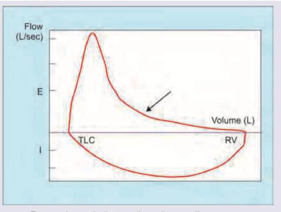
The following flow volume curve indicates:

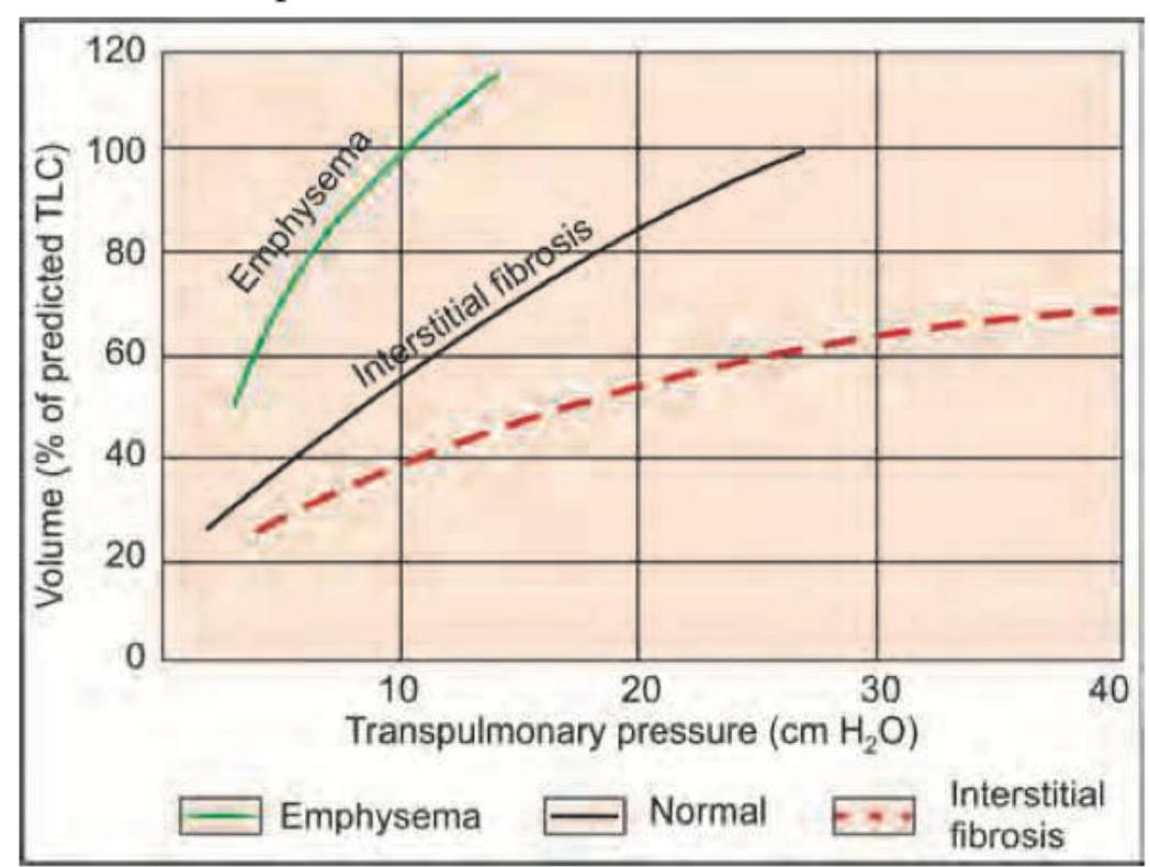
The curve shown is shifted downwards and to the right by which of the following?

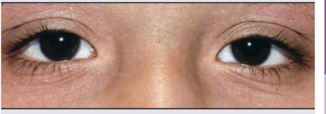
Which of the following is seen in the image?

Explanation: ***Nasal polyp***- This clinical scenario is classic for **Aspirin-Exacerbated Respiratory Disease (AERD)**, also known as **Samter's Triad**, a condition characterized by three key components.- The three components of Samter's Triad are **asthma** (wheezing/breathlessness), chronic rhinosinusitis with **nasal polyps**, and sensitivity to aspirin/NSAIDs [1].*IgE release*- **Aspirin sensitivity** is a pseudoallergic reaction driven by abnormal metabolism of **arachidonic acid**, specifically involving the COX-1 pathway, and is thus typically **non-IgE mediated**.- This pathway disturbance leads to the overproduction of bronchoconstrictive **leukotrienes** (LTC4, LTD4, LTE4), which are the primary mediators of the reaction.*Extrinsic asthma*- **Extrinsic asthma** refers to allergic asthma, which relies on **IgE-mediated Type I hypersensitivity** reactions triggered by environmental allergens.- AERD is classified as a form of **intrinsic (non-allergic) asthma** because the trigger mechanism is pharmacological/metabolic rather than immunological (allergen-specific IgE) [1].*Drug interaction*- The adverse reaction seen here is an **idiosyncratic drug hypersensitivity reaction** stemming from the patient's underlying disease (AERD), not interaction with another drug.- A true **drug interaction** occurs when one drug alters the absorption, distribution, metabolism, or excretion of another drug, which is irrelevant to leukotriene overproduction.
Explanation: ### Idiopathic pulmonary fibrosis (IPF) - The combination of progressive dyspnea, **digital clubbing**, and bilateral basal **fine inspiratory crackles (Velcro crackles)** is highly suggestive of IPF [1]. - HRCT findings of **subpleural reticular opacities** and **honeycombing** predominantly in the lower lobes represent the definitive usual interstitial pneumonia (**UIP**) pattern [1]. - There is a strong association with cigarette smoking, and the disease typically presents in older adults, often after age 50 [1]. ### Chronic obstructive pulmonary disease (COPD) - COPD is characterized by an **obstructive pattern** on PFTs (FEV1/FVC ratio <0.7), which contradicts the patient's normal ratio of 0.85. - HRCT in COPD primarily shows **centrilobular emphysema** or parenchymal destruction, rather than the severe reticulation and honeycombing described here. ### Hypersensitivity pneumonitis (HP) - Chronic HP often results in fibrosis that preferentially spares the **subpleural region** and typically lacks the classic, profound basal, subpleural **honeycombing** defining UIP [2]. - The typical HRCT findings involve ground-glass opacities, air trapping, and centrilobular nodules, often with a mid-to-upper lung predominance [2]. ### Sarcoidosis - Sarcoidosis most commonly presents with **bilateral hilar lymphadenopathy** and **upper lobe predominant** micronodules or fibrosis. - **Digital clubbing** is rare in sarcoidosis, and the disease pattern usually does not mimic the classic subpleural, basal predominant **UIP/honeycombing** seen here.
Explanation: ***Air trapping with hyperinflation of lungs*** - The FEV1/FVC ratio is **65% (below 70%)**, confirming an **obstructive lung disease** (e.g., COPD/emphysema) due to irreversible airflow limitation [2]. - An RV/TLC ratio of **141% (above 120%)** signifies **hyperinflation** (increased **Residual Volume** and **Total Lung Capacity**), which is the direct result of air trapping in the distal airspaces due to airway obstruction [1]. ***Decreased lung compliance*** - Decreased compliance is characteristic of **restrictive lung diseases** (e.g., pulmonary fibrosis), where the lungs are stiff and difficult to inflate. - In obstructive diseases like emphysema, the underlying pathophysiology often involves **destruction of alveolar walls**, which *increases* lung compliance. ***Restrictive lung disease*** - Restrictive diseases are characterized by a **normal or increased FEV1/FVC ratio** and decreased lung volumes (low FVC and TLC), which contradicts the PFT findings (low FEV1/FVC) [3]. - The main physiological problem is difficulty *expanding* the lungs, not difficulty *expelling* air. ***Increased lung compliance*** - While increased compliance is seen in pure emphysema, the PFT findings specifically point to the *consequences* of airway obstruction, which is **air trapping** and subsequent **hyperinflation** (increased RV/TLC ratio) [1]. - Describing the underlying pathophysiology as only 'increased lung compliance' is incomplete, as the most immediate and defining physiological result of severe obstruction leading to dyspnea is the high residual volume due to trapped air.
Explanation: ***Start L.A.B.A + inhaled corticosteroids*** - Nocturnal symptoms and night-time awakening indicate persistent or **partially controlled asthma**, necessitating a step-up in therapy (Step 3 or higher treatment based on GINA guidelines) [1]. - The preferred step-up involves adding a daily **low-dose inhaled corticosteroid (ICS)**, often combined with a **Long-Acting Beta Agonist (LABA)**, to address underlying inflammation and provide long-acting bronchodilation [1]. *Increase the dose of salbutamol* - **Salbutamol is a Short-Acting Beta Agonist (SABA)**, used for quick relief (rescue medication), not for controlling chronic inflammation or preventing nocturnal symptoms. - Increasing SABA use without adding anti-inflammatory medication (ICS) is an inappropriate strategy for managing **persistent asthma** and signals poor control. *Add theophylline* - Theophylline is a non-selective phosphodiesterase inhibitor, which is a **less preferred add-on therapy** (Step 4/5) due to its narrow therapeutic index and significant risk of **adverse effects** (e.g., arrhythmias, seizures). - This option is generally reserved for patients who remain symptomatic despite adequate ICS/LABA combination therapy. *Add Montelukast* - Montelukast (a **leukotriene receptor antagonist**) is an optional, less potent add-on therapy typically considered if symptoms persist despite ICS use or if the patient has significant **allergic rhinitis**. - It is **less effective** than adding an ICS-LABA combination when stepping up treatment for poorly controlled persistent asthma.
Explanation: ***Chronic respiratory acidosis*** - The high **pCO2 (60 mmHg)** indicates primary respiratory acidosis, while the relatively stable pH (**7.32** is mildly acidotic) implies significant renal compensation (elevated **HCO3-**) [1]. - This compensated state, where the pH is buffered despite chronic hypercapnia, is characteristic of a long-standing disease like **COPD** [1], [3]. *Acute respiratory acidosis* - If the acidosis were acute, there would be insufficient time for the kidneys to retain bicarbonate, resulting in a **more severe acidemia** (pH typically <7.25) for a pCO2 of 60 mmHg [2]. - An acute picture is less typical in a patient with stable COPD, which is inherently a **chronic condition** [1]. *Metabolic acidosis* - Metabolic acidosis is defined by a primary reduction in **HCO3-**, leading to a low pH [2]. - In this case, the acidemia is driven by the primary elevation of **pCO2** (hypercapnia), not a decrease in bicarbonate. *Metabolic alkalosis* - This condition is characterized by a primary increase in **HCO3-**, leading to an elevated pH (**alkalemia**). - Since the patient's pH is low (**acidemic**), and the primary driving force is CO2 retention, this diagnosis is incorrect.
Explanation: ***CTPA*** - This is the **gold standard** for diagnosing acute pulmonary embolism (PE) by visualizing the filling defects in the pulmonary arteries [2]. - Given the high clinical suspicion (post-operative status, classical symptoms, elevated D-dimer) and hemodynamic instability, rapid confirmation with **CTPA** is essential for determining the need for treatments like thrombolysis [2], [3]. *V/Q scan* - **V/Q scans** are generally reserved for patients where CTPA is contraindicated, such as in severe renal failure or certain allergies, and are typically not performed in unstable patients [2]. - It frequently yields an intermediate probability result, which is less conclusive than a CTPA, especially when a definitive, rapid diagnosis is needed due to **hemodynamic compromise**. *ECG* - **ECG** is a vital part of the initial assessment to rule out conditions like acute myocardial infarction and to check for signs of right heart strain common in massive PE (e.g., S1Q3T3 pattern) [1]. - Although crucial for risk stratification, it is only a **supportive test** and is not the definitive diagnostic tool for confirming the presence of PE. *CXR* - The **CXR** is primarily useful for excluding other pulmonary pathologies that mimic PE, such as pneumonia or pneumothorax, and is often normal in the setting of acute PE [1]. - Findings associated with PE (like **Westermark's sign** or **Hampton hump**) are often subtle, non-specific, and lack the sensitivity required to confirm the diagnosis [1].
Explanation: ***Laba plus inhalation steroids*** - In a patient with persistent symptoms (nocturnal exacerbations) despite using a SABA (**salbutamol**) and a SAMA (**ipratropium**), the next step is to initiate or step up therapy by adding a long-term controller medication, which is typically a combination of **Inhaled Corticosteroids (ICS)** and a **Long-Acting Beta-Agonist (LABA)** [1]. - This combination is crucial for controlling airway inflammation and providing prolonged bronchodilation, effectively managing nocturnal symptoms [1], [2]. *Oral corticosteroids* - **Oral corticosteroids** are reserved for acute, severe exacerbations, short courses for bridging, or in cases of very severe asthma refractory to high-dose inhaled therapy [3]. - They are not the standard next step for managing persistent nocturnal symptoms in a patient previously stable on SABA/SAMA. *Montelukast* - **Montelukast** (a leukotriene receptor antagonist) is generally considered a less potent controller than the ICS/LABA combination and is often used as an add-on therapy or for specific phenotypes like aspirin-exacerbated respiratory disease (AERD). - Its efficacy in independently controlling moderate-to-severe persistent asthma, characterized by nocturnal symptoms, is lower than that of ICS/LABA [2]. *Increase the dose of salbutamol* - **Salbutamol** is a **Short-Acting Beta-Agonist (SABA)**, used primarily as a reliever for acute symptoms, not as a long-term controller [2], [4]. - Increasing its dose or frequency significantly suggests reliance on relief medication, which indicates poorly controlled asthma and can increase the risk of adverse cardiac side effects like **tachycardia**.
Explanation: ***Parenchymal obstructive airway disease*** - The flow-volume loop shows a **reduced peak expiratory flow** and a **concave slope** of the expiratory limb, characteristic of **airflow obstruction**. - This pattern, particularly with an increased RV and TLC, is typical for conditions like **emphysema** or **chronic bronchitis**, where there is loss of elastic recoil or airway narrowing. *Restrictive defect* - A restrictive defect would typically show a **narrower loop** with reduced lung volumes (TLC, RV), but the shape of the expiratory curve would be more normal or proportionally scaled down, without the prominent concavity. - The primary issue in restrictive diseases is reduced lung compliance, not airway obstruction, so flow rates might be preserved or even increased relative to lung volume. *Extrathoracic obstruction* - Extrathoracic obstruction (e.g., vocal cord dysfunction, goiter compressing the trachea) primarily affects the **inspiratory limb** of the flow-volume loop, causing **flattening of inspiration**. - The expiratory limb in extrathoracic obstruction is typically preserved or much less affected than inspiration. *Intrathoracic obstruction* - **Fixed intrathoracic obstruction** (e.g., tracheal stenosis) causes **flattening of both inspiratory and expiratory limbs**. - A **variable intrathoracic obstruction** (e.g., tracheomalacia) specifically causes **flattening of the expiratory limb** but does not typically result in the marked concavity seen in parenchymal obstructive disease.
Explanation: ***Interstitial fibrosis*** - Interstitial fibrosis causes the lung to become **stiffer** and less compliant, requiring a **greater transpulmonary pressure** to achieve a given volume. This shifts the pressure-volume curve to the **right and downwards**. - The fibrosis leads to increased **elastic recoil**, making it harder for the lungs to expand, thus reducing lung volumes like **Total Lung Capacity (TLC)**. *Emphysema* - Emphysema is characterized by increased lung compliance due to the destruction of **elastic fibers**, which shifts the pressure-volume curve to the **left and upwards**. - This condition results in **air trapping** and increased lung volumes at a given transpulmonary pressure. *Pulmonary edema* - Pulmonary edema can **reduce lung compliance** due to fluid accumulation in the interstitial spaces and alveoli, which would shift the curve to the right and downwards. - However, the primary clinical presentation and **pathophysiology typically focus on impaired gas exchange** and fluid overload rather than a fundamental change in static lung mechanics reflected by chronic fibrosis. *Bronchitis* - Chronic bronchitis is primarily an **obstructive disease** characterized by inflammation and mucus production in the airways, leading to airflow limitation. - While it can indirectly affect lung mechanics due to air trapping and hyperinflation, it primarily impacts dynamic airflow and is **not typically represented by a direct shift** of the static pressure-volume curve in the manner shown for restrictive diseases.
Explanation: **Mongoloid slant** * The image clearly depicts an **upward and outward slant of the palpebral fissures**, where the outer canthus is higher than the inner canthus. This is characteristic of a mongoloid slant. * This feature is often associated with certain genetic conditions like **Down syndrome**, but can also be a normal variant in individuals of East Asian descent. *Antimongoloid slant* * An antimongoloid slant would present with a **downward and outward slant of the palpebral fissures**, where the outer canthus is lower than the inner canthus. * This is characteristic of conditions like **Treacher Collins syndrome** or **Noonan syndrome**, which is not depicted here. *Dennie-Morgan lines* * **Dennie-Morgan lines** are folds or creases under the lower eyelid, often associated with **atopic dermatitis** or allergies. They are not the primary feature shown in the image, which focuses on the slant of the eyes. * While subtle lower lid creasing might be present, it's not the defining characteristic of the image and is not the same as the specific eye slant. *Blaschko lines* * **Blaschko lines** are invisible lines of normal cell development in the skin that become apparent in various linear skin conditions, such as **linear nevus sebaceous**. * They are **not related to ocular morphology or the appearance of the eyes** themselves, but rather to patterns of skin lesions.
Obstructive Airway Diseases (Asthma, COPD)
Practice Questions
Interstitial Lung Diseases
Practice Questions
Pulmonary Infections
Practice Questions
Pulmonary Vascular Diseases
Practice Questions
Pleural Diseases
Practice Questions
Sleep-Disordered Breathing
Practice Questions
Respiratory Failure
Practice Questions
Mediastinal Disorders
Practice Questions
Occupational Lung Diseases
Practice Questions
Pulmonary Function Testing
Practice Questions
Bronchiectasis and Cystic Fibrosis
Practice Questions
Lung Cancer Approach
Practice Questions
Get full access to all questions, explanations, and performance tracking.
Start For Free