
Pulmonology — MCQs

On this page
Upper lobe bronchiectasis is seen in which disease?
Which is the most common organ involved in sarcoidosis
Samters triad is seen in patients with ?
A male patient presents to the emergency department. The arterial blood gas report is as follows: pH, 7.2; pCO2, 81 mmHg; and HCO3, 40 meq/L. Which of the following is the most likely diagnosis?
A patient presents to you with fever, night sweats, ptosis, and bilateral facial nerve palsy. Investigations showed leukocytosis and bilateral hilar lymphadenopathy. Which of the following is the most likely diagnosis?
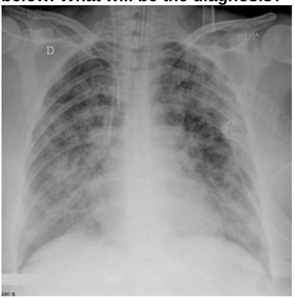
A patient with a known case of acute pancreatitis develops breathlessness and bilateral basal crepitations on day 4. What is the most likely diagnosis based on the chest radiography image?
A patient with a history of carcinoma of the bladder presents with dyspnoea, clinical signs of deep vein thrombosis (DVT), and tachycardia. Based on the Wells score for pulmonary embolism, what is the risk classification for this patient?
Oxygen therapy may not be useful in
What does a decreased FEV1/FVC ratio typically indicate in pulmonary function tests?
Which of the following is least likely to be associated with allergic bronchopulmonary aspergillosis (ABPA)?
Practice by Chapter
Obstructive Airway Diseases (Asthma, COPD)
Practice Questions
Interstitial Lung Diseases
Practice Questions
Pulmonary Infections
Practice Questions
Pulmonary Vascular Diseases
Practice Questions
Pleural Diseases
Practice Questions
Sleep-Disordered Breathing
Practice Questions
Respiratory Failure
Practice Questions
Mediastinal Disorders
Practice Questions
Occupational Lung Diseases
Practice Questions
Pulmonary Function Testing
Practice Questions
Bronchiectasis and Cystic Fibrosis
Practice Questions
Lung Cancer Approach
Practice Questions
Want unlimited practice?
Get full access to all questions, explanations, and performance tracking.
Scan to download app