
Pulmonology — MCQs

On this page
A patient developed breathlessness and chest pain, on second postoperative after a total hip replacement. Echo-cardiography showed right ventricular dilatation and tricuspid regurgitation. What is the most likely diagnosis.
Uveoparotid fever is seen in:
Most common extra-oral cause of Halitosis is?
Which of the following is least likely to be associated with hemothorax?
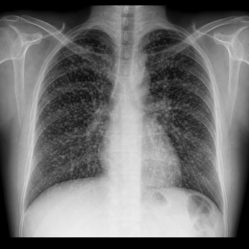
A 45-year-old crane operator at a construction site with pre-existing seropositive rheumatoid arthritis complains of progressive difficulty in breathing. On examination - Rheumatoid nodules are also present. Chest X-ray was performed. What is the diagnosis?
All are features of Silico-tuberculosis except:
Risk of thromboembolism is highest with:
For doing ABG, which of the following is used?
Patient with clinical signs of DVT had tachycardia and history of bladder cancer. According to modified Well's scoring, the probability of pulmonary embolism would be :
A female patient with clinical symptoms of systemic sclerosis presents with shortness of breath and bilateral basal rales. Her chest X-ray showed reticular opacities in bilateral basal fields. What is the next best step?
Practice by Chapter
Obstructive Airway Diseases (Asthma, COPD)
Practice Questions
Interstitial Lung Diseases
Practice Questions
Pulmonary Infections
Practice Questions
Pulmonary Vascular Diseases
Practice Questions
Pleural Diseases
Practice Questions
Sleep-Disordered Breathing
Practice Questions
Respiratory Failure
Practice Questions
Mediastinal Disorders
Practice Questions
Occupational Lung Diseases
Practice Questions
Pulmonary Function Testing
Practice Questions
Bronchiectasis and Cystic Fibrosis
Practice Questions
Lung Cancer Approach
Practice Questions
Want unlimited practice?
Get full access to all questions, explanations, and performance tracking.
Scan to download app