
Pulmonology — MCQs

On this page
A previously healthy 18-year-old high school student suddenly develops left-sided pleuritic chest pain and dyspnea. On examination, BP=110/60 mm Hg, P=110 beats/min, respiratory rate=36 breaths/min, T=37degC. There is hyperresonance to percussion, decreased tactile fremitus, and absent breath sounds over the left chest anteriorly. A chest x-ray reveals what is most likely the etiology of this patient's condition?
What is true about intrinsic asthma?
Which of the following is NOT true about aspirin-sensitive asthma?
A 74-year-old man with a history of smoking notices blood in his chronic daily sputum production. He has no fever or chills, but has lost 10 lb in the past 6 months. On examination, he has bilateral expiratory wheezes, and his fingers are clubbed. There are no lymph nodes and the remaining examination is normal. A chest X-ray reveals a left hilar mass. Which of the following suggests that the tumor is a small cell lung cancer?
Which of the following would be the most reasonable step in the assessment of a patient with emphysema due to alpha-1 antitrypsin deficiency (ATD)?
What is the most common cause of preventable hospital death?
Which of the following conditions is most sensitively detected by D-dimer testing?
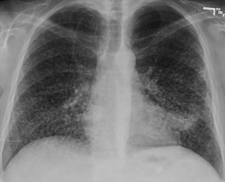
A 60-year-old man with a 40-pack-year smoking history and 20 years of work as a construction worker presents with shortness of breath and occasional blood-streaked sputum. His ECG shows lateral wall ischemia. What is the likely cause of the findings on his chest x-ray?
A 28-year-old bank employee presents with infertility and oligospermia. He reports a history of recurrent pancreatitis and chronic cough since childhood, with evident digital clubbing. Which test is most likely to reveal the cause of his chronic lung disease?
Dyspnoea at rest is which grade?
Practice by Chapter
Obstructive Airway Diseases (Asthma, COPD)
Practice Questions
Interstitial Lung Diseases
Practice Questions
Pulmonary Infections
Practice Questions
Pulmonary Vascular Diseases
Practice Questions
Pleural Diseases
Practice Questions
Sleep-Disordered Breathing
Practice Questions
Respiratory Failure
Practice Questions
Mediastinal Disorders
Practice Questions
Occupational Lung Diseases
Practice Questions
Pulmonary Function Testing
Practice Questions
Bronchiectasis and Cystic Fibrosis
Practice Questions
Lung Cancer Approach
Practice Questions
Want unlimited practice?
Get full access to all questions, explanations, and performance tracking.
Scan to download app