
Pulmonology — MCQs

On this page
A 58-year-old female smoker with end-stage chronic obstructive pulmonary disease and osteoarthritis, who is on ipratropium bromide, albuterol inhalers, and hydrocodone-acetaminophen, presents with respiratory distress for 2 days. She has increased thick, yellow sputum production, low-grade fever, and increasing confusion. On examination, she is mildly obtunded but arousable, with a blood pressure of 160/100 mmHg, pulse of 115/min, respiratory rate of 30/min, and O2 saturation of 84% on her usual 3 L/min nasal cannula oxygen. She is using accessory muscles to breathe, has diffuse wheezing and rhonchi bilaterally, a prolonged expiratory phase, distant but regular heart sounds, and no peripheral edema. Arterial blood gases (ABGs) on arrival are: pH 7.20, PO2 70 mmHg, PCO2 65 mmHg, calculated HCO3 29 mEq/L. Electrolytes are: Na 140 mEq/L, K 5.1 mEq/L, HCO3 29 mEq/L, Cl 100 mEq/L, BUN 20 mg/dL, creatinine 1.5 mg/dL, glucose 89 mg/dL. After prompt initiation of noninvasive positive pressure ventilation (Bi-pap), blood cultures, toxicology screen, intravenous fluids, and IV antibiotics, consider the patient's metabolic situation. Which of the following best describes the acid-base condition and its etiology?
A patient with acute pancreatitis develops sudden onset breathlessness with a CVP < 15 mm Hg. The chest X-ray shows bilateral infiltrates. What is the most likely diagnosis?
A patient presents with elevated JVP on the right side and a positive Kussmaul sign. Superior Vena Cava (SVC) obstruction is suspected. In which of the following is SVC obstruction commonly seen?
A 30-year-old woman presents with fatigue, dark "tea color" urine, and yellowish eyes. Her past medical history includes myasthenia gravis treated with azathioprine and pyridostigmine. Laboratory findings are consistent with autoimmune hemolytic anemia. A chest X-ray shows an anterior mediastinal mass. What is the most likely diagnosis?
On spirometry, decreased FEV1, normal FVC, increased TLC, and decreased DLCO2 suggest which diagnosis?
A 65-year-old man presents with a chronic cough, which has recently changed character. Tuberculosis and other infectious causes have been ruled out. What should be the next investigation?
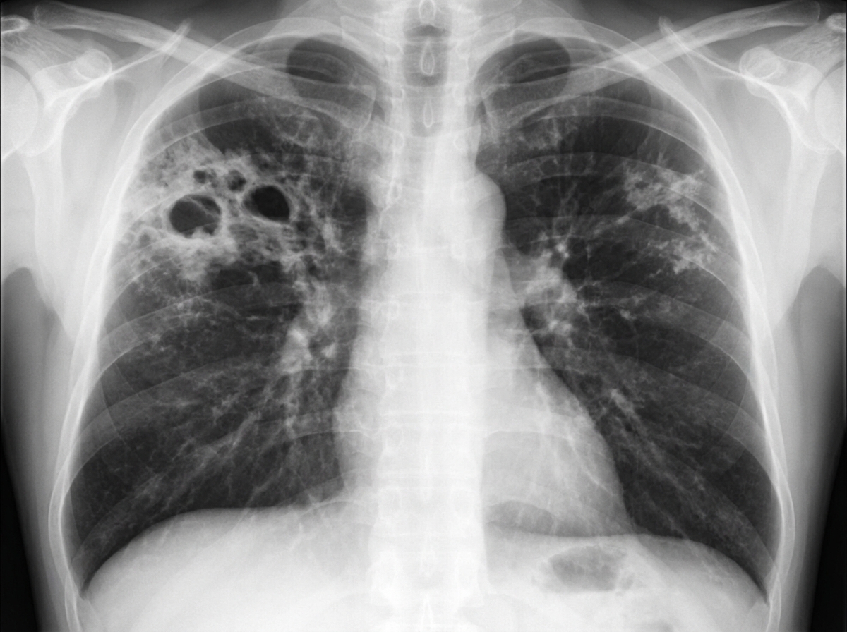
A 60-year-old man with a history of COPD and old TB presents with mild hemoptysis and chronic cough. He is HIV negative and has been ill for approximately 2 weeks. Vital signs: pulse 110 bpm; temperature 101°F; respirations 24/min; blood pressure 108/70 mm Hg. No skin lesions are noted. Laboratory data: Hb 14 g/dL; HCT 42%; WBCs 8.7/uL; BUN 24 mg/dL; creatinine 0.8 mg/dL; sodium 131 mEq/L; potassium 4.3 mEq/L. Arterial blood gases on room air: pH 7.37; PCO2 43 mm Hg; PO2 87 mm Hg. Sputum tests reveal numerous AFB-positive organisms on smear. Spirometry shows an obstructive ventilatory impairment with marginal reversibility. Chest X-ray is provided. Among the choices listed, what is the most likely diagnosis?
What is the most common cause of fungal infection in a known case of bronchial asthma with eosinophilia?
Which one of the following is not likely to be associated with pulmonary fibrosis?
A 32-year-old patient presents with complaints of difficulty in breathing, chest pain, and increased respiratory rate. Pulmonary thromboembolism is suspected. Which investigation is most useful in acute pulmonary thromboembolism?
Practice by Chapter
Obstructive Airway Diseases (Asthma, COPD)
Practice Questions
Interstitial Lung Diseases
Practice Questions
Pulmonary Infections
Practice Questions
Pulmonary Vascular Diseases
Practice Questions
Pleural Diseases
Practice Questions
Sleep-Disordered Breathing
Practice Questions
Respiratory Failure
Practice Questions
Mediastinal Disorders
Practice Questions
Occupational Lung Diseases
Practice Questions
Pulmonary Function Testing
Practice Questions
Bronchiectasis and Cystic Fibrosis
Practice Questions
Lung Cancer Approach
Practice Questions
Want unlimited practice?
Get full access to all questions, explanations, and performance tracking.
Scan to download app