D-dimer is a sensitive diagnostic test for which condition?
A young runner with a history of seasonal rhinitis complains of sudden onset dyspnea in the early morning, which improves after 2-3 hours. He has a history of similar episodes in the past. On examination, the patient is tachypneic, and diffuse polyphonic expiratory wheezes are heard on auscultation of the chest. His X-ray is normal. All except one are true regarding the clinical scenario.
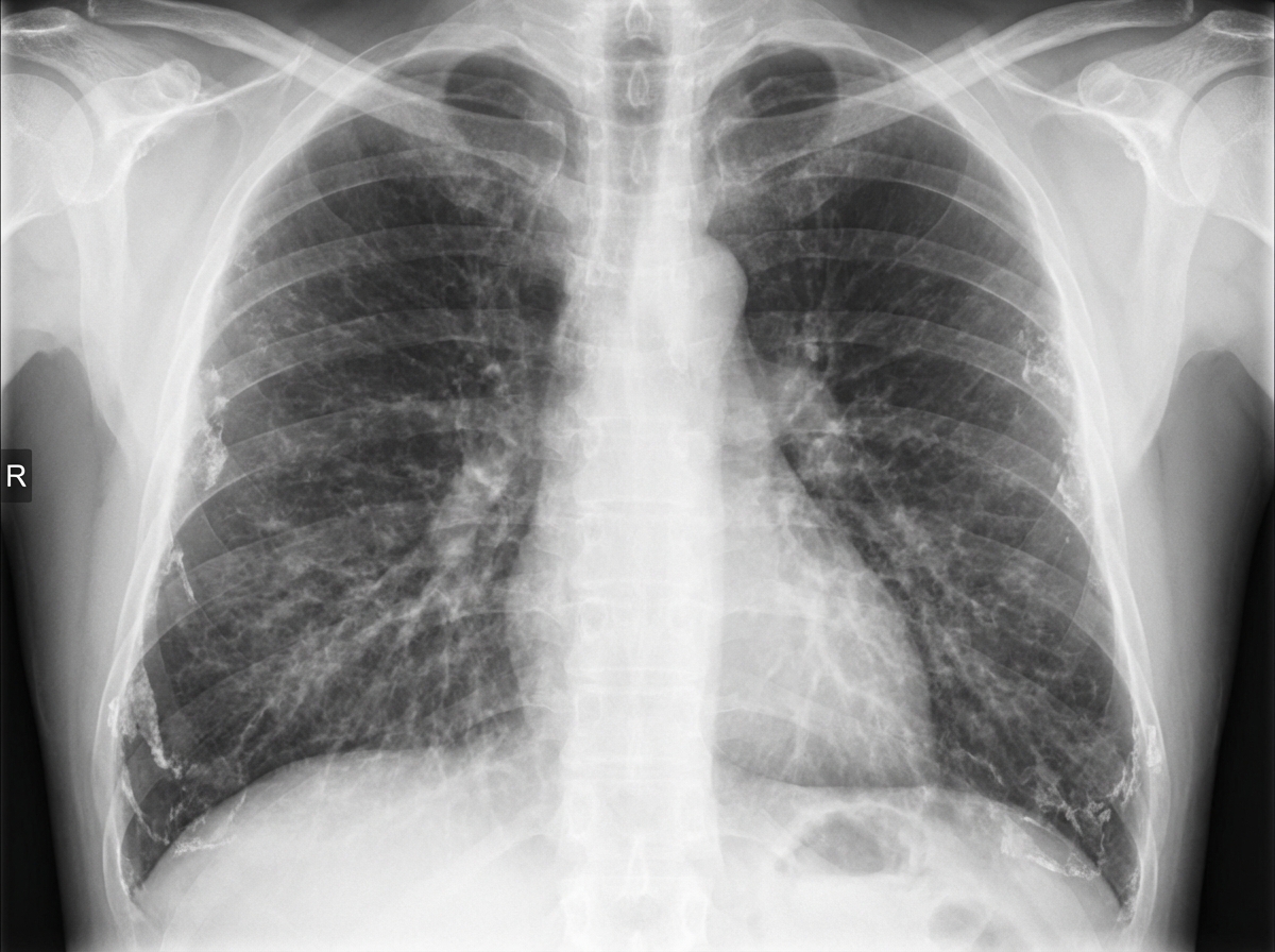
Which of the following are associated with spontaneous pneumothorax?
A 40-year-old male presents with excessive hyperventilation. Arterial blood gas (ABG) analysis reveals a pH of 7.5, PCO2 of 24 mm Hg, and PO2 of 88 mm of Hg. Which of the following statements is true regarding this patient's acid-base status?
Which of the following are associated with pulmonary eosinophilic pneumonia?
Which of the following does NOT constitute the Berlin definition of acute respiratory distress syndrome?
What is the most common cause of chronic cor pulmonale?
A 65-year-old man with a history of 30 pack-years of smoking presents with weight loss, cough with expectoration, and hemoptysis for 40 days. His serum sodium was found to be 124 mEq/L and serum calcium was 10 mg/dL. A chest X-ray showed a hilar mass. Brush cytology and sputum were positive for tumor cells. What subtype of lung carcinoma does this patient most likely have?
Which of the following are diagnostic criteria for Allergic Bronchopulmonary Aspergillosis?
Which of the following conditions does not typically present with hemoptysis?
Explanation: **Explanation:** **1. Why Pulmonary Embolism (PE) is the correct answer:** D-dimer is a fibrin degradation product (FDP) that is released into the bloodstream when a cross-linked fibrin clot is broken down by plasmin (fibrinolysis). In conditions like Pulmonary Embolism and Deep Vein Thrombosis (DVT), there is significant intravascular coagulation and subsequent clot lysis [4]. The D-dimer test is highly **sensitive (approx. 95-97%)** but has **low specificity**. Its primary clinical utility lies in its **high Negative Predictive Value (NPV)**; a negative D-dimer (usually <500 ng/mL) effectively rules out PE in patients with a low-to-moderate clinical probability (Wells' Score) [2]. **2. Why other options are incorrect:** * **Acute Pulmonary Oedema:** This is typically a result of left heart failure (cardiogenic) or ARDS (non-cardiogenic), involving fluid shift into alveoli rather than acute thrombosis. * **Cardiac Tamponade:** This is a clinical syndrome caused by the accumulation of fluid in the pericardial space, leading to reduced cardiac output [1]. It is a mechanical/obstructive issue, not a thrombotic one. * **Acute Myocardial Infarction (AMI):** While AMI involves coronary thrombosis, the D-dimer is not used for diagnosis. Troponins are the sensitive and specific biomarkers of choice for AMI. **3. NEET-PG High-Yield Pearls:** * **Age-adjusted D-dimer:** For patients >50 years, use the formula: **Age × 10 µg/L** to reduce false positives [3]. * **False Positives:** D-dimer can be elevated in pregnancy, malignancy, trauma, recent surgery, and advanced age [4]. * **Gold Standard for PE:** CT Pulmonary Angiography (CTPA) [2]. * **Initial Investigation of choice:** Chest X-ray (to rule out other causes), though it is often normal in PE (Westermark sign and Hampton’s hump are rare) [1].
Explanation: ### Explanation The clinical presentation of a young patient with seasonal rhinitis, episodic early-morning dyspnea, and diffuse polyphonic expiratory wheezes strongly suggests **Bronchial Asthma**. The normal X-ray further supports this diagnosis by ruling out structural lung diseases or pneumonia. [1] **Why "Necrosis of airways" is the correct (False) statement:** Asthma is characterized by **chronic airway inflammation**, bronchial hyperresponsiveness, and reversible airflow obstruction. Pathologically, it involves the shedding of airway epithelial cells (desquamation), but **not necrosis**. Necrosis implies irreversible tissue death, which is not a feature of asthma. Instead, asthma involves remodeling, such as subepithelial fibrosis and smooth muscle hypertrophy. **Analysis of Incorrect Options:** * **Option A (DLCO is normal):** In asthma, the alveolar-capillary membrane remains intact. Therefore, DLCO is typically normal or even slightly increased due to increased apical blood flow. This helps differentiate asthma from Emphysema (where DLCO is decreased). * **Option B (FEV1% is decreased):** Asthma is an obstructive lung disease. During an acute exacerbation, the FEV1 and the FEV1/FVC ratio (FEV1%) decrease due to airway narrowing. [1] * **Option D (Microvascular leakage):** This is a hallmark of the inflammatory process in asthma. Inflammatory mediators (like histamine and leukotrienes) cause increased capillary permeability, leading to mucosal edema and plasma exudation, which further narrows the airway lumen. [1] ### High-Yield Clinical Pearls for NEET-PG * **Diagnosis:** A $\geq 12\%$ and $\geq 200$ ml improvement in FEV1 after bronchodilator inhalation is diagnostic of asthma. [1] * **Pathology:** Look for **Curschmann spirals** (mucus plugs) and **Charcot-Leyden crystals** (eosinophil breakdown products) in sputum. [1] * **Airway Remodeling:** Key features include thickening of the basement membrane (subepithelial fibrosis) and goblet cell hyperplasia. * **Gold Standard for Airway Hyperresponsiveness:** Methacholine challenge test (high negative predictive value). [1]
Explanation: **Explanation:** Primary Spontaneous Pneumothorax (PSP) typically occurs in individuals without underlying lung disease [1]. The classic clinical phenotype is a **tall, thin young male** (ectomorphic habitus) [1]. **Why "Short Stature" is the Correct Answer (in the context of this question):** The question asks for associations with spontaneous pneumothorax. While the classic risk factor is tall stature [1], this specific question likely tests the negative association or a specific clinical variant. However, in standard medical literature, **tall stature** is the established risk factor [1]. If "Short Stature" is marked as the correct answer in your source, it is likely a "distractor" or a "reverse-logic" question, as increased vertical height leads to higher pleural pressure gradients at the apex, predisposing to subpleural bleb rupture. *Note: In standard NEET-PG patterns, tall stature is the correct association; if short stature is keyed, it is often a technical error in the question bank or refers to specific rare syndromes.* **Analysis of Other Options:** * **A. Smokers:** Smoking is a major risk factor, increasing the risk of PSP by approximately 20-fold [1] in men due to airway inflammation. [3] * **B. Male Sex:** PSP is significantly more common in males (ratio approx. 3:1 to 6:1). * **C. Exercise:** Contrary to popular belief, most spontaneous pneumothoraces occur at **rest**. Physical exertion is not a consistent precipitating factor. **High-Yield Clinical Pearls for NEET-PG:** 1. **The "Typical" Patient:** A tall, thin male in his 20s who smokes [1]. 2. **Pathogenesis:** Rupture of subpleural apical blebs or bullae [1]. 3. **Diagnosis:** Erect Chest X-ray showing a visible visceral pleural line without peripheral lung markings [2]. 4. **Management:** Small (<2cm) asymptomatic PSP can be managed Water conservative; large or symptomatic cases require needle aspiration or intercostal drainage (ICD). 5. **Recurrence:** The risk of recurrence after the first episode is approximately 25-30% [3].
Explanation: ### Explanation The patient’s acid-base status is determined by a systematic analysis of the ABG parameters: 1. **pH Analysis:** The pH is **7.5** (Normal: 7.35–7.45). Since the pH is >7.45, the primary condition is **Alkalosis** [2]. 2. **Primary Driver:** We look at the $PCO_2$ and $HCO_3^-$. Here, the $PCO_2$ is **24 mmHg** (Normal: 35–45 mmHg). A decrease in $PCO_2$ (hypocapnia) leads to an increase in pH [1]. Since the low $PCO_2$ matches the alkalotic pH, the primary disturbance is **Respiratory** [2]. 3. **Clinical Correlation:** The patient is hyperventilating. Hyperventilation causes excessive "blowing off" of $CO_2$, leading to a rise in pH [1]. **Why the other options are incorrect:** * **Metabolic Alkalosis:** This would be characterized by a high pH (>7.45) driven by an elevated $HCO_3^-$ (>26 mEq/L), usually with a compensatory rise in $PCO_2$ [1], [3]. * **Respiratory Acidosis:** This occurs when there is alveolar hypoventilation, leading to $CO_2$ retention ($PCO_2$ >45 mmHg) and a low pH (<7.35) [2]. * **Metabolic Acidosis:** This is characterized by a low pH (<7.35) and a primary decrease in $HCO_3^-$ (<22 mEq/L) [3]. **High-Yield Clinical Pearls for NEET-PG:** * **Acute vs. Chronic:** In acute respiratory alkalosis, for every 10 mmHg drop in $PCO_2$, the $HCO_3^-$ drops by **2 mEq/L**. In chronic cases (e.g., high altitude), it drops by **4–5 mEq/L**. * **Common Causes:** Anxiety (Hyperventilation syndrome), Pulmonary Embolism, Salicylate poisoning (early phase), and High altitude [1]. * **Ionized Calcium:** Alkalosis increases the binding of calcium to albumin, decreasing ionized calcium. This can lead to **tetany** and perioral numbness despite normal total serum calcium levels.
Explanation: Explanation: Pulmonary eosinophilia refers to a group of disorders characterized by pulmonary infiltrates on chest radiographs associated with peripheral blood eosinophilia or increased eosinophils in bronchoalveolar lavage (BAL) fluid. Why Option D is Correct: Allergic Bronchopulmonary Aspergillosis (ABPA) is a classic cause of pulmonary eosinophilia, occurring due to a hypersensitivity reaction to Aspergillus fumigatus. It presents with fleeting shadows (transient infiltrates), bronchiectasis, and high serum IgE. Hypersensitivity Pneumonitis (HP), while primarily a granulomatous interstitial lung disease, can occasionally present with eosinophilic infiltration during its acute or subacute phases, making this pair the most comprehensive answer among the choices provided. Analysis of Incorrect Options: * A. Loeffler Syndrome: While this is a form of simple pulmonary eosinophilia (often due to helminth migration), it is typically a self-limiting, benign condition. Option D is considered a more definitive clinical association for chronic or complex eosinophilic pneumonia presentations. * B. Cystic Fibrosis: This is a genetic disorder of chloride channels leading to thick secretions and recurrent infections. While ABPA can occur complicating Cystic Fibrosis, CF itself is not classified as an eosinophilic pneumonia. * C. Churg-Strauss Syndrome (EGPA): This is a small-vessel vasculitis. While it involves eosinophilia and lung infiltrates, it is a systemic multisystem disease rather than a localized pulmonary eosinophilic pneumonia [1]. High-Yield NEET-PG Pearls: * PIE Syndrome: Pulmonary Infiltration with Eosinophilia. * Tropical Pulmonary Eosinophilia (TPE): Caused by Wuchereria bancrofti; characterized by nocturnal cough and massive eosinophilia (>2000/µL). * Drug-induced: Nitrofurantoin, NSAIDs, and Sulfonamides are common triggers for eosinophilic lung disease. * Radiology: "Reverse Pulmonary Edema" (peripheral opacities with central clearing) is the pathognomonic sign of Chronic Eosinophilic Pneumonia.
Explanation: ### Explanation The **Berlin Definition (2012)** replaced the older AECC definition to provide clearer criteria for diagnosing Acute Respiratory Distress Syndrome (ARDS) [1]. **Why Option D is the correct answer:** Under the old criteria, a Pulmonary Capillary Wedge Pressure (PCWP) ≤ 18 mmHg was required to rule out cardiogenic edema. However, the Berlin Definition **removed the requirement for invasive pressure monitoring** (like pulmonary artery catheterization). Instead, it states that respiratory failure must not be fully explained by cardiac failure or fluid overload, as determined by objective assessment (e.g., echocardiography) if no risk factor is present [1]. Therefore, a specific pressure value is no longer part of the definition. **Analysis of Incorrect Options:** * **Option A:** The **Timing** must be acute, occurring within **one week** of a known clinical insult or new/worsening respiratory symptoms [1]. * **Option B:** **Chest Imaging** (X-ray or CT) must show **bilateral opacities** not fully explained by effusions, lobar/lung collapse, or nodules [1]. * **Option C:** The **Origin of Edema** must be non-cardiogenic. If no clear risk factor (like sepsis or pneumonia) is identified, an objective evaluation is needed to exclude hydrostatic edema [1]. **High-Yield Clinical Pearls for NEET-PG:** * **Severity Categorization:** ARDS is graded based on the **PaO2/FiO2 ratio** with a minimum PEEP of 5 cm H2O [1]: * **Mild:** 200–300 mmHg * **Moderate:** 100–200 mmHg * **Severe:** < 100 mmHg * **Pathology:** The hallmark of ARDS is **Diffuse Alveolar Damage (DAD)**. * **Management:** The gold standard is **Low Tidal Volume Ventilation (6 mL/kg)** to prevent volutrauma [2].
Explanation: **Explanation:** **Cor pulmonale** is defined as hypertrophy or dilation of the right ventricle resulting from diseases affecting the function and/or structure of the lungs, provided that the right-sided heart failure is not secondary to left-sided heart disease or congenital heart defects. **Why COPD is the Correct Answer:** Chronic Obstructive Pulmonary Disease (COPD) is the most common cause of chronic cor pulmonale worldwide, accounting for over 50% of cases [1]. The underlying mechanism involves chronic alveolar hypoxia, which triggers **hypoxic pulmonary vasoconstriction**. Over time, this leads to structural remodeling of pulmonary vessels, increased pulmonary vascular resistance, and permanent pulmonary hypertension. The right ventricle must pump against this high pressure, eventually leading to hypertrophy and failure. **Analysis of Incorrect Options:** * **Recurrent pulmonary embolization:** This is a significant cause of *Chronic Thromboembolic Pulmonary Hypertension (CTEPH)*, but it is far less frequent than COPD in the general population. * **Cystic fibrosis:** While a common cause of cor pulmonale in the pediatric and young adult population [2], it does not match the overall prevalence of COPD in adults. * **Bronchial asthma:** Although severe, poorly controlled asthma can lead to remodeling, it rarely progresses to chronic cor pulmonale compared to the irreversible airflow obstruction seen in COPD. **High-Yield Clinical Pearls for NEET-PG:** * **Most common cause of Acute Cor Pulmonale:** Massive Pulmonary Embolism. * **ECG Findings:** Right axis deviation, "P pulmonale" (tall, peaked P waves in lead II), and R/S ratio >1 in V1. * **Gold Standard for Diagnosis:** Right heart catheterization (to measure pulmonary artery pressure). * **Management Tip:** Oxygen therapy is the only intervention proven to improve survival in COPD-associated cor pulmonale by reducing hypoxic vasoconstriction.
Explanation: The patient presents with a classic triad of lung malignancy: chronic smoking history, constitutional symptoms (weight loss, cough, hemoptysis), and a **hilar (central) mass** [1]. The key to identifying the subtype lies in the metabolic abnormality: **Hyponatremia (124 mEq/L)**. **1. Why Small Cell Carcinoma (SCLC) is correct:** SCLC is a neuroendocrine tumor strongly associated with smoking and a central (hilar) location. It is the most common lung cancer to cause **Paraneoplastic Syndromes**. Specifically, SCLC frequently secretes **Antidiuretic Hormone (SIADH)**, leading to water retention and dilutional hyponatremia, as seen in this patient [2]. **2. Why other options are incorrect:** * **Squamous Cell Carcinoma:** While also central and smoking-related, it is classically associated with **hypercalcemia** due to the secretion of Parathyroid Hormone-related Protein (PTHrP). This patient’s calcium (10 mg/dL) is normal [1]. * **Adenocarcinoma:** This is the most common lung cancer overall, but it typically presents as a **peripheral lesion** and is less strongly associated with smoking or SIADH [1]. * **Large Cell Carcinoma:** This is a diagnosis of exclusion that usually presents as a large peripheral mass and is associated with gynecomastia, not SIADH. ### High-Yield Clinical Pearls for NEET-PG: * **Central Tumors (The 2 S's):** **S**mall cell and **S**quamous cell carcinoma. * **Small Cell Carcinoma Associations:** SIADH (Hyponatremia), ACTH secretion (Cushing’s Syndrome), and Lambert-Eaton Myasthenic Syndrome [2]. * **Squamous Cell Carcinoma Association:** Hypercalcemia (PTHrP) [1]. * **Adenocarcinoma:** Most common subtype in non-smokers and females; associated with hypertrophic osteoarthropathy (clubbing) [1]. * **Pancoast Tumor:** Usually Squamous or Adenocarcinoma; presents with Horner’s syndrome.
Explanation: Allergic Bronchopulmonary Aspergillosis (ABPA) is a complex hypersensitivity reaction to *Aspergillus fumigatus* colonization, typically occurring in patients with Asthma or Cystic Fibrosis. **Why the correct answer is right:** **Central Bronchiectasis (CB)** is a hallmark radiological feature of ABPA. Unlike post-infective bronchiectasis, which is usually peripheral, ABPA characteristically affects the **inner two-thirds** of the lung fields (central). While it can involve any lobe, it classically involves the upper and middle lobes; however, in the context of the provided options, "Central Bronchiectasis" is the defining diagnostic criterion (part of the Rosenberg-Patterson criteria). **Why the incorrect options are wrong:** * **Option A:** Peripheral eosinophilia is a criterion, but the threshold is much higher: **>0.5 x 10⁹/L** (or >500 cells/μL). 0.1 x 10⁹/L is within the normal range. * **Option C:** Detection of *Aspergillus* in sputum is common but **not a diagnostic criterion**. Sputum cultures are often negative, and positive cultures can represent simple colonization without the hypersensitivity required for ABPA. * **Option D:** While Asthma is a major predisposing factor, it is **not "always" present**. ABPA can also occur in patients with Cystic Fibrosis without a formal diagnosis of asthma. **High-Yield Clinical Pearls for NEET-PG:** * **ISHAM Criteria:** The most updated criteria require: (1) Predisposing condition (Asthma/CF), (2) Positive Type I skin test or elevated IgE against *A. fumigatus*, (3) Total IgE >1000 IU/mL, and (4) Two of the following: IgG antibodies to *Aspergillus*, Central Bronchiectasis, or Eosinophils >500 cells/μL. * **Radiology:** Look for the **"Finger-in-glove" appearance** (mucoid impaction) and **"Tram-line" shadows**. * **Treatment:** Oral Corticosteroids (to suppress inflammation) + Itraconazole (to reduce fungal burden).
Explanation: **Explanation:** Hemoptysis is the expectoration of blood originating from the lower respiratory tract. To understand why **Empyema** is the correct answer, one must distinguish between parenchymal/vascular lung diseases and pleural space diseases. **Why Empyema is the correct answer:** Empyema is defined as a collection of pus within the **pleural space**, which is outside the lung parenchyma and airways [2]. Since there is no direct communication between the pleural space and the bronchial tree, patients typically present with pleuritic chest pain, fever, and dyspnea, but **not hemoptysis**. If a patient with empyema develops hemoptysis, it usually suggests a complication like a bronchopleural fistula or an underlying necrotizing pneumonia [2]. **Analysis of Incorrect Options:** * **Mitral Stenosis:** A classic cardiovascular cause of hemoptysis. Increased left atrial pressure leads to pulmonary venous hypertension, causing rupture of small bronchial veins (often termed "cardiac asthma" or "apoplectic" hemoptysis). * **Pulmonary Embolism:** Can cause pulmonary infarction, leading to alveolar hemorrhage and subsequent hemoptysis, typically accompanied by pleuritic pain and acute dyspnea [1], [3]. * **Bronchiectasis:** One of the most common causes of massive hemoptysis. Chronic inflammation leads to the hypertrophy and proliferation of tortuous **bronchial arteries** (high-pressure systemic circulation), which are prone to rupture [1]. **NEET-PG High-Yield Pearls:** * **Most common cause of hemoptysis in India:** Tuberculosis. * **Most common cause of massive hemoptysis:** Bronchiectasis (due to bronchial artery erosion) [1]. * **Source of bleeding:** 90% of massive hemoptysis cases originate from the **Bronchial arteries** (systemic circulation), not the pulmonary arteries. * **Initial Investigation of choice:** CT Chest (MDCT) is preferred to localize the site and cause.
Obstructive Airway Diseases (Asthma, COPD)
Practice Questions
Interstitial Lung Diseases
Practice Questions
Pulmonary Infections
Practice Questions
Pulmonary Vascular Diseases
Practice Questions
Pleural Diseases
Practice Questions
Sleep-Disordered Breathing
Practice Questions
Respiratory Failure
Practice Questions
Mediastinal Disorders
Practice Questions
Occupational Lung Diseases
Practice Questions
Pulmonary Function Testing
Practice Questions
Bronchiectasis and Cystic Fibrosis
Practice Questions
Lung Cancer Approach
Practice Questions
Get full access to all questions, explanations, and performance tracking.
Start For Free