
Pulmonology — MCQs

On this page
A patient presents with non-productive cough, hemoptysis, and grade III clubbing. Chest X-ray reveals an upper left zone lesion. What is the likely cause?
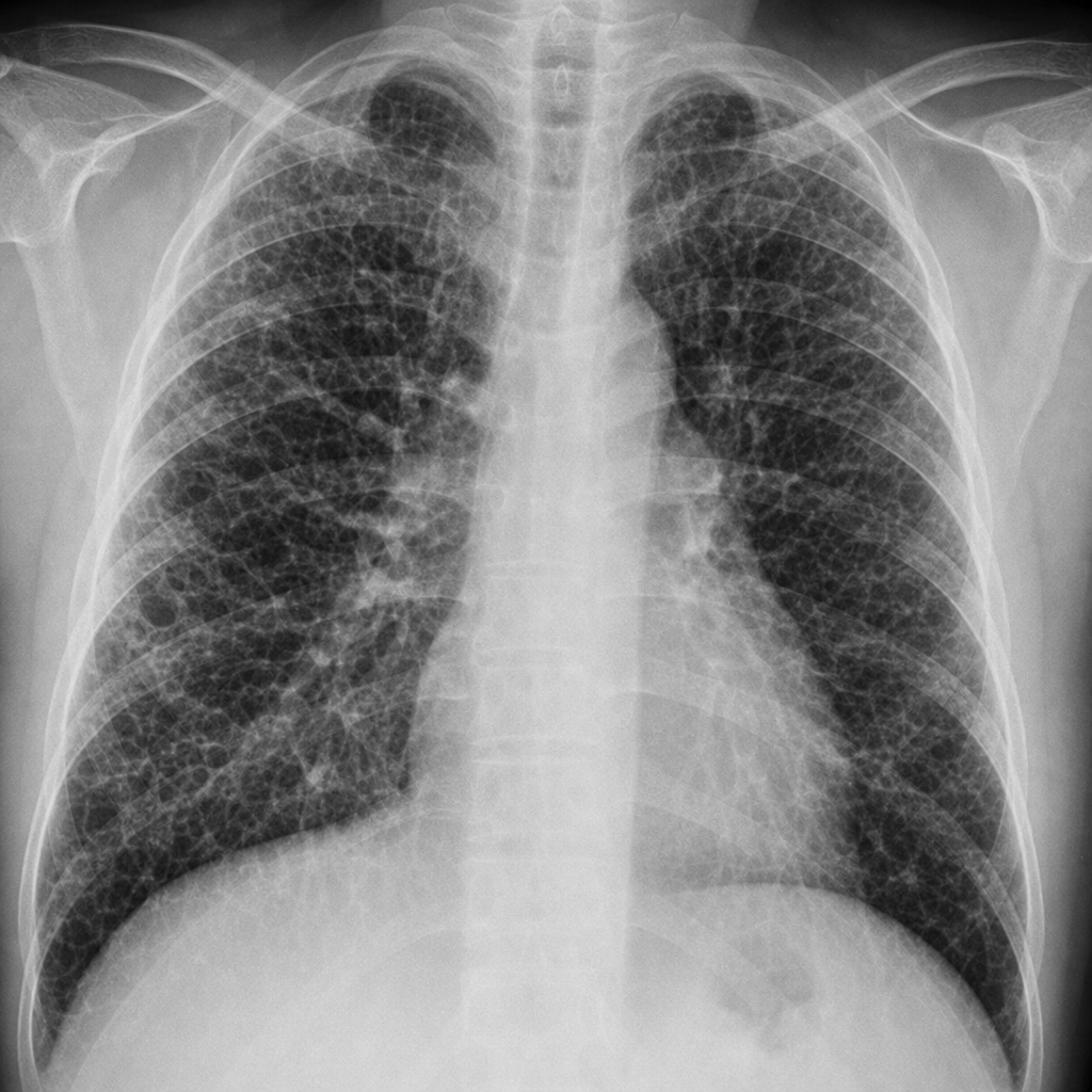
A patient presented with bilateral hilar lymphadenopathy and a negative Mantoux test. What is the most likely diagnosis?
A 30-year-old female patient complains of breathlessness for the past 5 months. Her pulmonary function tests report is as follows:
All of the following test results can exclude pulmonary embolism, EXCEPT:
A 64-year-old woman is found to have a left-sided pleural effusion on chest x-ray. Analysis of the pleural fluid reveals a ratio of concentration of total protein in pleural fluid to serum of 0.38, a lactate dehydrogenase (LDH) level of 125 IU, and a ratio of LDH concentration in pleural fluid to serum of 0.46. Which of the following disorders is most likely in this patient?
A 50-year-old woman presents with a 4-week history of fever, shortness of breath, and dry cough. She reports that her chest feels 'tight.' The patient is a pigeon fancier. Blood tests show leukocytosis and neutrophilia, an elevated erythrocyte sedimentation rate, and increased levels of immunoglobulins and C-reactive protein. A lung biopsy reveals poorly formed granulomas composed of epithelioid macrophages and multinucleated giant cells. Which of the following is the most likely diagnosis?
An adolescent girl presents with symmetrical, red, tender swellings in her shins and arthralgia. X-ray reveals hilar and paratracheal lymphadenopathy. She is clinically suspected to have sarcoidosis. What is the next step in the diagnosis?
The Bode index is used in which type of transplantation?
In COPD, which of the following statements is true?
A 45-year-old patient on methotrexate develops respiratory symptoms. What is the most likely diagnosis?
Practice by Chapter
Obstructive Airway Diseases (Asthma, COPD)
Practice Questions
Interstitial Lung Diseases
Practice Questions
Pulmonary Infections
Practice Questions
Pulmonary Vascular Diseases
Practice Questions
Pleural Diseases
Practice Questions
Sleep-Disordered Breathing
Practice Questions
Respiratory Failure
Practice Questions
Mediastinal Disorders
Practice Questions
Occupational Lung Diseases
Practice Questions
Pulmonary Function Testing
Practice Questions
Bronchiectasis and Cystic Fibrosis
Practice Questions
Lung Cancer Approach
Practice Questions
Want unlimited practice?
Get full access to all questions, explanations, and performance tracking.
Scan to download app