
Pulmonology — MCQs

On this page
A 65-year-old man presented with hemoptysis and stage 3 clubbing. What is the probable diagnosis?
Which of the following is NOT a consequence of obstructive sleep apnea?
A 74-year-old woman, in otherwise good health, sustained a leg injury 2 days ago and has been bedridden since the accident. Two hours ago, she became delirious. On physical examination, her temperature is 99 F, blood pressure is 120/70 mmHg, heart rate is 110, and respiratory rate is 32. Pulse oximetry shows an oxygen saturation of 80%, and a chest x-ray film is normal. Which of the following is the most likely diagnosis?
Which one of the following parameters is consistent with pleural transudate?
Tuberculosis is associated with which of the following conditions?
A 30-year-old male presents with bilateral lymphadenitis characterized by non-caseating granulomas on biopsy. What is the most likely diagnosis?
A 55-year-old man presents with increasing shortness of breath and dry cough for several years. He has a significant smoking history and daily alcohol consumption. He experiences severe breathlessness with minimal exertion, prolonged expiration with wheezing, a barrel chest, hyperresonance on percussion, digital clubbing, facial plethora, and leg edema. Chest X-ray shows hyperinflation, flattened diaphragms, and increased retrosternal air space. What is the most likely diagnosis?
Which of the following is the cause of sudden and unexpected onset of dyspnea at rest?
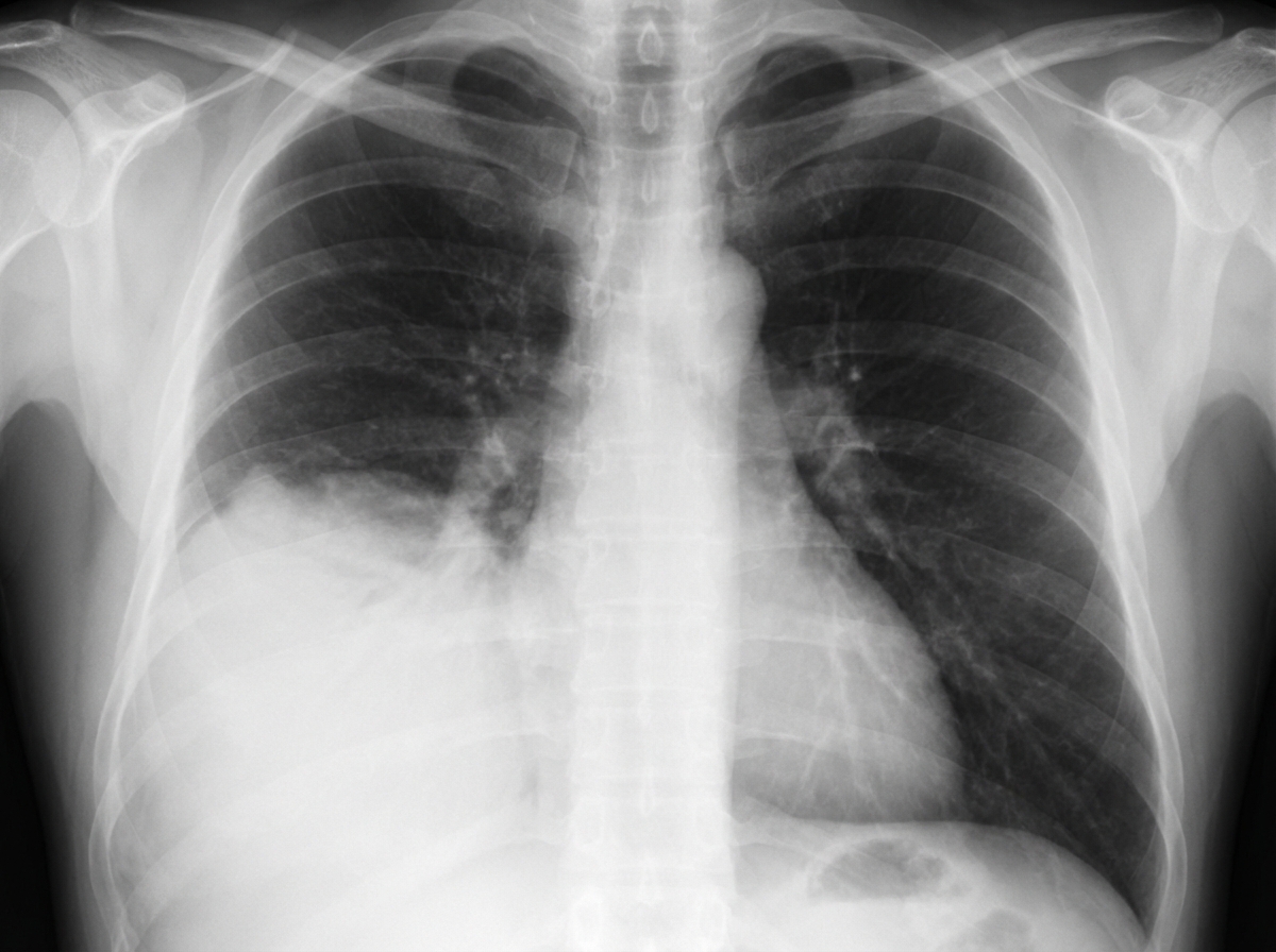
A 40-year-old male presented to the OPD with fever and breathlessness. His X-ray is shown below. What is the most probable diagnosis?
All of the following statements about silicosis are true, EXCEPT:
Practice by Chapter
Obstructive Airway Diseases (Asthma, COPD)
Practice Questions
Interstitial Lung Diseases
Practice Questions
Pulmonary Infections
Practice Questions
Pulmonary Vascular Diseases
Practice Questions
Pleural Diseases
Practice Questions
Sleep-Disordered Breathing
Practice Questions
Respiratory Failure
Practice Questions
Mediastinal Disorders
Practice Questions
Occupational Lung Diseases
Practice Questions
Pulmonary Function Testing
Practice Questions
Bronchiectasis and Cystic Fibrosis
Practice Questions
Lung Cancer Approach
Practice Questions
Want unlimited practice?
Get full access to all questions, explanations, and performance tracking.
Scan to download app