What is the T stage of a 2.5cm lung carcinoma, not involving the pleura?
A 45-year-old male reports several years of asbestos exposure while working in the construction industry. He reports smoking 2 packs of cigarettes per day for over 20 years. Smoking and asbestos exposure increase the incidence of which of the following diseases?
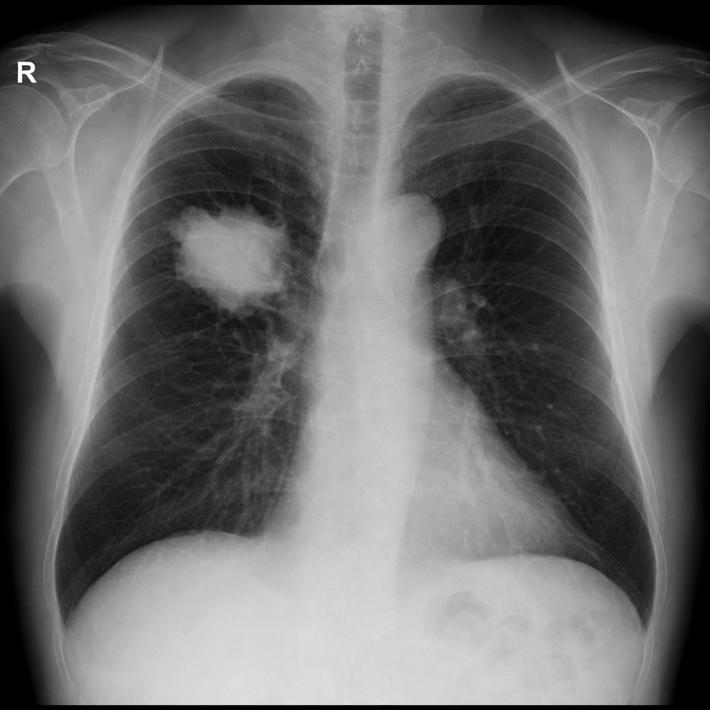
A 70-year-old smoker presents with dyspnea and weight loss. CXR shows a mass in the right lung. What is the next step?
What finding during surgery can change the staging of a tumor from Stage I to Stage II in a patient with a history of lung cancer?
Which type of lung cancer is most commonly found in non-smokers?
What is the most appropriate next step in management for a patient with a Stage III ovarian cancer with partial response to platinum-based chemotherapy?
Which of the following is the MOST characteristic feature of Eaton-Lambert syndrome?
Which of the following is a characteristic of bronchoalveolar carcinoma?
Most common symptom of lung carcinoma is
Stereotactic Radiotherapy is used in:
Explanation: ***T1c*** - A **2.5 cm lung carcinoma** without pleural involvement falls into the T1 category [1]. - According to the **TNM staging system (8th edition)** for lung cancer, a tumor between **2-3 cm is classified as T1c** [1]. *T1a* - This classification is reserved for tumors that are **1 cm or less** in greatest dimension. - The given tumor size of **2.5 cm is larger** than the T1a criteria. *T2* - A T2 tumor is generally defined by a size greater than **3 cm but less than or equal to 5 cm**, or has specific features like visceral pleural invasion or involvement of the main bronchus regardless of distance from the carina [1]. - Our tumor is **only 2.5 cm** and does not involve the pleura, excluding T2. *T1b* - This category applies to tumors that are **greater than 1 cm but equal to or less than 2 cm** in greatest dimension. - The 2.5 cm tumor size exceeds the criteria for **T1b**. **References:** [1] Kumar V, Abbas AK, et al.. Robbins and Cotran Pathologic Basis of Disease. 9th ed. The Lung, pp. 721-725.
Explanation: ***Bronchogenic carcinoma*** - **Smoking** is the leading cause of **bronchogenic carcinoma**, and **asbestos exposure** significantly *multiplies* its risk, rather than simply adding to it. - This synergistic effect means that smokers exposed to asbestos have a **much higher incidence** of lung cancer compared to those with either exposure alone. *Emphysema* - Primarily linked to **smoking** and chronic exposure to irritants, but asbestos exposure does not significantly increase its incidence. - While both smoking and asbestos can cause pulmonary issues, their primary mechanisms for emphysema are distinct. *Malignant pulmonary mesothelioma* - **Malignant mesothelioma** is strongly associated with **asbestos exposure**, but its incidence is *not significantly increased* by smoking. - Smoking is a risk factor for lung cancer, but not a primary risk factor for mesothelioma itself. *Multiple myeloma* - This is a **hematologic malignancy** (cancer of plasma cells) and has no established link with either **smoking** or **asbestos exposure**. - Its risk factors are largely genetic and related to other environmental factors, but not directly linked to respiratory toxins. *Chronic bronchitis* - **Chronic bronchitis** is primarily caused by **smoking** and exposure to environmental pollutants. - While asbestos exposure can cause lung damage, it doesn't directly or significantly increase the incidence of chronic bronchitis.
Explanation: ***CT-guided biopsy*** - A definitive diagnosis of a suspected lung mass, especially in a patient with a history of smoking and weight loss, requires **histological confirmation**. [1] - **CT-guided biopsy** is often the most direct and least invasive method to obtain tissue from a peripheral lung mass for pathological examination. [2] *Bronchoscopy* - While useful for diagnosing central airway lesions, **bronchoscopy** may not reach a peripheral lung mass, making tissue sampling difficult or impossible. [3] - It could be considered if the mass were closer to the main bronchi or if the patient had symptoms suggesting endobronchial involvement. [4] *MRI* - **MRI** is not the primary imaging modality for initial evaluation of a lung mass; **CT scans** provide better anatomical detail of the lungs and chest wall. [2] - It might be used for staging purposes, especially for evaluating brain metastases or chest wall invasion, but not for initial diagnosis of the lung mass itself. *Sputum cytology* - **Sputum cytology** has low sensitivity for diagnosing lung cancer, especially for peripheral lesions, as cancer cells may not be exfoliated into the sputum. - A negative result from sputum cytology does not rule out lung cancer, and a more invasive procedure would still be needed for diagnosis.
Explanation: ***Positive bronchial lymph nodes*** - The presence of **positive bronchial lymph nodes** (N1) indicates regional lymph node involvement, necessitating an upgrade to Stage II from Stage I [1]. - This finding is significant in lung cancer staging, suggesting metastasis beyond the primary tumor. *Tumor at the carina* - A tumor at the **carina** may imply local invasion but does not specifically relate to lymph node involvement for upgrading the stage. - This would indicate a more advanced tumor stage only if it invaded adjacent structures directly. *Involvement of the chest wall* - Chest wall involvement typically refers to **direct extension of the tumor** and might upgrade the stage to III, not II. - The initial staging focused on **nodal involvement**, which is not indicated in this case. *Small cell histology* - Small cell carcinoma, while aggressive and often systemic, does not correspond with this staging system based on **N classification**. - It also usually presents with different clinical features and patterns compared to non-small cell lung cancers. **References:** [1] Kumar V, Abbas AK, et al.. Robbins and Cotran Pathologic Basis of Disease. 9th ed. The Lung, p. 725.
Explanation: ***Adenocarcinoma*** - It is the most common type of lung cancer among **non-smokers**, often associated with **lung scarring** and **asbestos exposure**. - Typically presents in the **peripheral** regions of the lungs and has a **glandular** pattern on histology. *Squamous cell carcinoma* - More commonly associated with **smoking** [1] and usually arises in the **central parts** of the lungs. - Known for cavitary lesions and often linked to chronic **lung disease** rather than non-smokers. *None of the above* - This oes not provide a definitive lung cancer type, which is **not relevant** when adenocarcinoma is the correct answer. - Fails to recognize the significant evidence linking adenocarcinoma as the predominant type in non-smokers. *Oat cell carcinoma* - This represents **small cell lung cancer**, which is strongly linked to **smoking** rather than being common in non-smokers [1]. - Often presents with **metastatic lesions** and is more aggressive, unlike adenocarcinoma.
Explanation: ***Perform surgery (Interval Debulking Surgery)*** - In **Stage III ovarian cancer**, after an initial partial response to **platinum-based chemotherapy**, **interval debulking surgery** is the standard next step to remove residual disease. - This approach aims to reduce tumor burden to an optimal level (< 1 cm residual disease), which has been shown to improve overall survival in multiple trials (EORTC 55971, GOG-152). - Performed after 3-4 cycles of neoadjuvant chemotherapy when the patient has demonstrated response and is medically fit for surgery. *Bevacizumab* - **Bevacizumab** is an **anti-angiogenic agent** used in ovarian cancer, typically as part of frontline maintenance therapy or for recurrent disease, not as the immediate next step after partial response to primary chemotherapy when surgery is feasible. - While it can be incorporated into maintenance treatment post-surgery, it's not the primary next step after partial response when interval debulking surgery is indicated. *Switch to radiotherapy* - **Radiotherapy** has a limited role in the primary treatment of advanced ovarian cancer due to its widespread peritoneal nature. - It is sometimes used for localized recurrence or symptom palliation, but not as a standard next step after partial response to chemotherapy in Stage III disease. *Continue regimen* - Continuing the same regimen after only a **partial response** is generally not the most effective strategy when further tumor reduction via surgery is possible. - The goal in advanced ovarian cancer is **maximal cytoreduction**, and if residual disease is present after neoadjuvant chemotherapy, interval debulking surgery is preferred over continued chemotherapy alone.
Explanation: ***Repeated electrical stimulation enhances muscle power in it.*** - A hallmark feature of **Lambert-Eaton Myasthenic Syndrome (LEMS)** is the **potentiation of muscle strength** with repeated or high-frequency nerve stimulation [2]. - This is due to the disease pathophysiology where repeated stimulation allows the accumulation of **intracellular calcium**, leading to increased acetylcholine release at the neuromuscular junction. *Neostigmine is not effective for this syndrome.* - While it's largely true that **acetylcholinesterase inhibitors** like neostigmine are less effective in LEMS compared to myasthenia gravis, they can still provide some minor symptomatic relief [1]. - Therefore, stating it's *not effective* might be an oversimplification, and it's not the *most characteristic* feature. *It is commonly associated with small cell lung cancer.* - Although LEMS is frequently a **paraneoplastic syndrome** linked to **small cell lung cancer (SCLC)**, this association is a cause/etiology, not a direct characteristic feature of the neuromuscular dysfunction itself [1], [2]. - Approximately 50-60% of LEMS cases are paraneoplastic, with SCLC being the most common underlying malignancy [2]. *It can affect the ocular muscles.* - **Ocular muscle involvement** (e.g., ptosis, diplopia) is a prominent and often initial symptom in **myasthenia gravis** [2]. - In LEMS, ocular muscle weakness is **much less common** and typically mild, if present, distinguishing it from myasthenia gravis.
Explanation: ***Adenocarcinoma*** - Bronchoalveolar carcinoma is classified as a subtype of **adenocarcinoma**, specifically presenting as non-small cell lung cancer (NSCLC) [1]. - It is characterized by **lepidic growth pattern** in the alveolar structures, which preserves the architecture of the lung parenchyma. *Stromal invasion with desmoplasia* - Typically, bronchoalveolar carcinoma shows **minimal invasion**, contrasting with the extensive desmoplastic reaction seen in other types of lung cancer. - This type is more about growth patterns than typical invasive features associated with stromal changes. *Grows along pre-existing anatomical structures* - While some lung tumors may grow along bronchi, bronchoalveolar carcinoma primarily **grows along alveolar surfaces** rather than conforming to anatomical structures. - This growth pattern leads to its distinct histological features, differing from the infiltrative patterns of other cancers. *Preservation of Alveolar structure* - Although bronchoalveolar carcinoma does preserve some architecture, stating it relies solely on this aspect is misleading as this does not comprehensively define the tumor. - Its distinction lies in its subtype classification as an **adenocarcinoma** rather than merely structural preservation [1]. **References:** [1] Cross SS. Underwood's Pathology: A Clinical Approach. 6th ed. Common Clinical Problems From Respiratory Tract Disease, pp. 335-336.
Explanation: ***Cough*** - **Chronic cough** is the most frequently reported symptom in patients with lung carcinoma, often progressing in severity or character. [1] - It results from irritation of the **bronchial tree** by the tumor or associated inflammation. [1] *Dyspnoea* - While common, **shortness of breath** typically occurs when the tumor significantly obstructs airways, causes pleural effusion, or spreads to lymphatics. [1] - It usually presents later in the disease progression compared to cough. *Weight loss* - **Unexplained weight loss** is a common systemic symptom of malignancy, including lung cancer, but is often a sign of advanced disease. [1] - This symptom is non-specific and can be associated with many chronic illnesses. *Chest pain* - **Chest pain** in lung carcinoma often indicates involvement of the pleura, chest wall, or mediastinum by the tumor. - It is a common symptom but is less frequent than cough as the initial presenting complaint.
Explanation: ***Inoperable Stage 1 Lung Tumor*** - **Stereotactic Radiotherapy (SRT)** is highly effective for **inoperable Stage 1 lung tumors** because it delivers high doses of radiation with extreme precision, maximizing tumor control while sparing surrounding healthy tissue. - The **precision** of SRT makes it an excellent option for localized, small tumors that cannot be surgically removed due to patient comorbidities or tumor location. *Lymphangitis Carcinomatosa* - **Lymphangitis carcinomatosa** involves diffuse infiltration of the pulmonary lymphatic system by cancer cells and is not amenable to localized radiation techniques like SRT. - Treatment for lymphangitis carcinomatosa typically involves **systemic therapy** such as chemotherapy or targeted therapy, rather than focal radiation. *Miliary Lung Metastasis* - **Miliary lung metastases** refer to widespread, small (~2-4mm) nodules throughout both lungs, indicating advanced systemic disease. - SRT is a **localized treatment** and therefore not appropriate for diffuse, multifocal disease like miliary metastases, which requires systemic treatment. *Tumor at the base of tongue with new Lymph Node enlargement* - A tumor at the **base of the tongue with new lymph node enlargement** indicates a larger disease burden, likely requiring a combination of **surgery, conventional radiation therapy, and/or chemotherapy**. - While radiation is a component of treatment for head and neck cancers, **SRT is typically reserved for smaller, well-defined lesions**, or in specific cases as a boost or for recurrences, not usually for initial treatment of a larger primary tumor with nodal involvement.
Get full access to all questions, explanations, and performance tracking.
Start For Free