Cystic fibrosis leads to defect in which of the following channels?
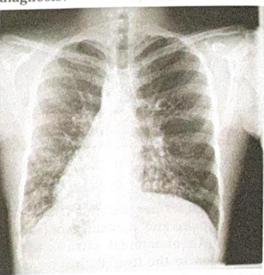
A 40-year-old man presented with repeated episodes of bronchospasm and hemoptysis. Chest X-ray revealed perihilar bronchiectasis. The most likely diagnosis is
In bronchiectasis, all of the following are seen except:
Ring sign with dilated bronchi on CXR is a feature of
Chloride level in sweat is used in the diagnosis of which disease?
Which of the following is not typically associated with cystic fibrosis?
A 3-year-old child is evaluated by a pediatrician for poor growth despite excessive food intake. The mother reports that the child's stools are bulky, foul-smelling, and difficult to flush because they float. Determination of which of the following would most likely be diagnostic in this case?
Investigation of choice in bronchiectasis?
A patient presents with recurrent lung infections, and the chest X-ray provided shows a characteristic finding. What is the most likely diagnosis?

Post-tubercular bronchiectasis is most commonly seen with
Explanation: ***Cl-*** - Cystic fibrosis is caused by a mutation in the **CFTR gene**, which encodes for the **Cystic Fibrosis Transmembrane Conductance Regulator protein**. - This protein functions primarily as a **chloride channel**, and its dysfunction leads to impaired chloride transport across epithelial cell membranes. *K+* - While potassium channels are crucial for many physiological processes, their primary dysfunction is **not directly linked to the pathogenesis of cystic fibrosis**. - Defects in potassium channels are associated with conditions like **long QT syndrome** or certain forms of epilepsy. *Ca2+* - **Calcium channels play a role in various cellular signaling pathways**, but their direct defect is not the underlying cause of cystic fibrosis. - Conditions like **Lambert-Eaton myasthenic syndrome** involve antibodies affecting presynaptic calcium channels. *Na+* - **Sodium channels are involved in maintaining membrane potential and fluid balance**, and while they interact with CFTR, their primary defect is not the cause of cystic fibrosis. - Dysregulation of sodium transport can occur secondary to CFTR dysfunction, leading to **dehydrated mucus**, but the initial defect is in chloride.
Explanation: ***Bronchopulmonary aspergillosis*** - **Allergic bronchopulmonary aspergillosis (ABPA)** is characterized by **bronchospasm**, recurrent fleeting pulmonary infiltrates, and **hemoptysis** in patients with asthma or cystic fibrosis. - The chest X-ray findings of **perihilar bronchiectasis** are a hallmark of ABPA, resulting from airway damage caused by the allergic response to *Aspergillus fumigatus*. *Sarcoidosis* - Sarcoidosis is a **granulomatous disease** that typically presents with non-caseating granulomas, often affecting the lungs, lymph nodes, eyes, and skin. [1] - While it can cause respiratory symptoms, **bronchiectasis** and **hemoptysis** are not typical primary features; more common findings include bilateral hilar lymphadenopathy and interstitial lung disease. [1] *Idiopathic pulmonary fibrosis* - Idiopathic pulmonary fibrosis (IPF) is a **chronic, progressive interstitial lung disease** characterized by progressive scarring of the lung tissue. [2] - While IPF causes respiratory symptoms like dyspnea and cough, it does not typically present with recurrent **bronchospasm**, **hemoptysis**, or **perihilar bronchiectasis**. [2] *Extrinsic allergic alveolitis* - Extrinsic allergic alveolitis, also known as **hypersensitivity pneumonitis**, is an immune-mediated lung disease caused by exposure to various inhaled antigens. - It typically presents with flu-like symptoms, cough, and dyspnea, and while it can cause interstitial lung disease, **bronchospasm** and **bronchiectasis** are not characteristic features.
Explanation: ***Wheezing*** - **Wheezing** is typically associated with conditions that cause airway narrowing, such as **asthma** or **COPD**. - While some patients with bronchiectasis might have concomitant airway obstruction, wheezing is **not a universal or characteristic symptom** of bronchiectasis itself, which primarily involves permanent dilation and destruction of the bronchi. *Clubbing* - **Clubbing** (thickening of the nail beds) is a common sign in patients with **chronic suppurative lung diseases** like bronchiectasis, indicating chronic hypoxia and inflammation [2]. - It results from the **persistent inflammation** and increased vascularity in the distal digits. *Haemoptysis* - **Haemoptysis**, or coughing up blood, is a frequent and sometimes severe complication of bronchiectasis due to the **fragile, hypervascular bronchial walls** that easily bleed [1], [2]. - The dilated and inflamed bronchi are highly prone to **vascular damage**, leading to varying degrees of bleeding. *Recurring bouts of cough* - **Chronic cough** productive of large amounts of purulent sputum is the **hallmark symptom** of bronchiectasis, resulting from chronic infection and inflammation [1]. - The impaired mucociliary clearance leads to **stagnant mucus** and recurrent infections, triggering persistent coughing.
Explanation: ***Bronchiectasis*** - The "ring sign" on a Chest X-Ray (CXR) is characteristic of **bronchiectasis**, representing the cross-sectional view of a **dilated bronchus**. - **Bronchial wall thickening** and dilation create this appearance, often accompanied by **"tram lines"** (parallel lines corresponding to longitudinal views of dilated bronchi). *Asthma* - CXR in asthma is often normal but may show signs of **hyperinflation** or **bronchial wall thickening** in severe cases. - It does not typically present with the "ring sign" of permanently dilated bronchi. *Bronchiolitis* - This condition primarily affects the **small airways (bronchioles)** and is common in infants. - CXR findings typically include **hyperinflation**, **peribronchial thickening**, and **atelectasis**, but not dilated bronchi forming ring signs. *Candidiasis* - Pulmonary candidiasis is a fungal infection that can cause various CXR findings like **infiltrates**, **nodules**, or **cavities**. - It does not typically lead to bronchial dilation or the characteristic "ring sign" seen in bronchiectasis.
Explanation: ***Cystic fibrosis*** - **Cystic fibrosis** is characterized by a defect in the **CFTR protein**, leading to impaired chloride transport in epithelial cells. - This defect results in abnormally high salt content in sweat, making the **sweat chloride test** the gold standard for diagnosis. *Phenylketonuria* - **Phenylketonuria (PKU)** is a metabolic disorder involving the inability to metabolize **phenylalanine**, leading to its accumulation in the blood. - Diagnosis is typically made via **newborn screening** using blood tests, not sweat chloride levels. *Gaucher's disease* - **Gaucher's disease** is a lysosomal storage disorder caused by a deficiency in the enzyme **beta-glucosidase**, leading to lipid accumulation. - Diagnosis involves enzyme assays from blood or tissue samples, or genetic testing, not sweat chloride analysis. *Osteogenesis imperfecta* - **Osteogenesis imperfecta** is a genetic disorder of **collagen synthesis**, primarily characterized by bone fragility, blue sclerae, and hearing loss. - Diagnosis is based on clinical features, imaging, and genetic testing, with no relevance to sweat chloride levels. *Tay-Sachs disease* - **Tay-Sachs disease** is a lysosomal storage disorder caused by deficiency of **hexosaminidase A**, leading to accumulation of GM2 ganglioside in neurons. - Diagnosis is made through **enzyme assay** or **genetic testing**, not sweat chloride measurement.
Explanation: ***Renal failure*** - **Renal failure** is not a typical manifestation or complication of **cystic fibrosis (CF)**. The CFTR protein primarily affects epithelial cells in organs like the lungs, pancreas, liver, and intestines, not the kidneys. - While CF patients may experience **electrolyte imbalances** due to excessive salt loss, this rarely progresses to **kidney failure** as a direct result of the disease itself. *Infertility* - **Infertility** is commonly associated with CF, particularly in males due to **congenital bilateral absence of the vas deferens (CBAVD)**, which is found in over 95% of male CF patients [1]. - In females, CF can cause **viscous cervical mucus** and nutritional deficiencies, leading to challenges with conception [1]. *Azoospermia* - **Azoospermia**, specifically **obstructive azoospermia**, is a direct consequence of **congenital bilateral absence of the vas deferens (CBAVD)**, which is highly prevalent in male CF patients [1]. - This condition prevents sperm from reaching the ejaculate, despite normal sperm production in the testes. *Nasal polyps* - **Nasal polyps** are a common feature in patients with **cystic fibrosis**, often leading to **chronic rhinosinusitis** and nasal obstruction. - This is due to the dysfunctional CFTR protein leading to abnormal mucus production and inflammation in the nasal passages [1].
Explanation: ***Na+ in sweat*** - This clinical presentation of **poor growth despite excessive food intake** (due to malabsorption) and **bulky, foul-smelling, floating stools** (steatorrhea) is highly suggestive of **cystic fibrosis (CF)**. - The **sweat test** is the diagnostic gold standard for CF. While the test measures both Na+ and Cl- in sweat, the **diagnostic criterion is based primarily on elevated chloride concentration** (Cl- ≥60 mmol/L). In CF, both sodium and chloride are elevated due to defective CFTR channel function. - This patient's symptoms of malabsorption result from **pancreatic insufficiency**, a common manifestation of CF. *Na+ in cerebrospinal fluid* - Measuring Na+ in cerebrospinal fluid is typically done to evaluate neurological conditions or electrolyte imbalances affecting the **central nervous system**, which is not indicated here. - There is no direct link between CSF Na+ levels and the malabsorption or growth failure described in this case. *Na+ in urine* - Urinary Na+ levels are used to assess **renal function** and overall **fluid and electrolyte balance**, and are relevant in conditions like **dehydration**, **kidney disease**, or **adrenal disorders**. - These tests are not primary diagnostic tools for the gastrointestinal and growth issues described, which point more towards a malabsorption syndrome. *Na+ in serum* - Serum Na+ levels reflect the body's overall **hydration status** and are crucial for diagnosing **hyponatremia** or **hypernatremia**. - While important for general medical assessment, serum Na+ levels do not specifically diagnose the underlying cause of malabsorption and steatorrhea seen in this child.
Explanation: ***HRCT*** - **High-resolution computed tomography (HRCT)** is the gold standard for diagnosing bronchiectasis, as it provides detailed images of the bronchial tree. - It effectively visualizes the characteristic **bronchial dilation**, **lack of bronchial tapering**, and **"signet-ring" appearance** of the airways. *MRI* - **Magnetic resonance imaging (MRI)** is generally not the primary imaging modality for bronchiectasis due to its lower spatial resolution compared to CT for lung parenchyma. - While it can provide functional information, it is not as effective in visualizing the characteristic anatomical changes of bronchiectasis. *Bronchoscopy* - **Bronchoscopy** is an invasive procedure primarily used to identify the cause of bronchiectasis (e.g., foreign body, infection, endobronchial obstruction) or for therapeutic lavage. - It is not the initial diagnostic investigation of choice for confirming the presence and extent of bronchiectasis itself. *Chest X-ray* - A **chest X-ray** may show non-specific findings such as increased bronchial wall opacity or tram-track lucencies, which are suggestive of bronchiectasis but not definitive. - It lacks the sensitivity and specificity of HRCT to confirm the diagnosis and delineate the extent of the disease.
Explanation: ***Kartagener syndrome*** - This syndrome is a subgroup of **primary ciliary dyskinesia** and is characterized by the triad of **situs inversus** (dextrocardia as seen on the chest X-ray), **chronic sinusitis**, and **bronchiectasis**. - Recurrent lung infections are a common presentation due to impaired mucociliary clearance leading to bronchiectasis. *Cystic fibrosis* - While cystic fibrosis does present with **recurrent lung infections** and **bronchiectasis**, it is not typically associated with **situs inversus** or other malformations of organ placement. - Diagnosis is usually supported by a **positive sweat chloride test** and genetic testing for CFTR mutations. *DiGeorge syndrome* - This syndrome is characterized by **T-cell immunodeficiency**, **hypocalcemia**, and **congenital heart defects**. - Recurrent infections in DiGeorge syndrome are due to immunodeficiency, not primarily due to impaired ciliary function or situs inversus. *Down syndrome* - Down syndrome is a chromosomal disorder associated with intellectual disability, distinctive facial features, and an increased risk of several health problems, including **congenital heart disease** and **immune dysfunction**. - Recurrent lung infections can occur due to weakened immune function or structural airway abnormalities, but it does not cause situs inversus or primary ciliary dyskinesia.
Explanation: ***Tuberculosis*** - **Tuberculosis (TB)**, particularly childhood TB, is a leading cause of post-infectious bronchiectasis, especially in regions with high TB prevalence [1]. - The inflammatory and destructive processes associated with TB infection in the lungs can lead to irreversible dilation and damage of the bronchi [1]. *Pertussis* - While **pertussis** can cause severe respiratory inflammation and chronic cough, it is a less common cause of widespread, irreversible bronchiectasis compared to tuberculosis [1]. - The damage caused by pertussis is typically more acute and less likely to lead to long-term structural changes like those seen in post-tubercular bronchiectasis. *Cystic fibrosis* - **Cystic fibrosis** is a genetic disorder that causes thick, sticky mucus to build up in the lungs, leading to chronic infections and bronchiectasis [1]. - However, post-tubercular bronchiectasis refers specifically to bronchiectasis developing *after* a tuberculosis infection, not as a primary genetic condition. *Kartagener syndrome* - **Kartagener syndrome** is a genetic disorder characterized by defects in ciliary function, leading to impaired mucociliary clearance and recurrent respiratory infections, which can result in bronchiectasis [1]. - Similar to cystic fibrosis, this is a primary genetic cause of bronchiectasis, distinct from bronchiectasis occurring as a sequela of tuberculosis.
Get full access to all questions, explanations, and performance tracking.
Start For Free