Which of the following are blood values of Iron Deficiency Anaemia ? 1. Serum iron is less than 30 mg/100 mL 2. Total iron binding capacity is less than 400 µg/mL 3. Percentage saturation is 10% or less 4. Serum ferritin is below 30 µg/mL Select the correct answer using the code given below :
Which of the following statements are correct regarding iron deficiency? 1. More common in older age group 2. More common in females of reproductive age 3. Associated with heavy menstrual bleeding 4. Commonly caused by chronic blood loss Select the correct answer using the code given below.
Which of the following statements are correct in respect of haemophilia? 1. It is hereditary bleeding disorder. 2. It has a higher incidence among Asians. 3. It affects females occasionally and mildly.
The following are the common features of Idiopathic Thrombocytopenic Purpura except
Priapism in a young male could occur because of
Umbilical cord blood stem cells are used to treat all the following diseases except :
Consider the following statements: Haemophilia A (haemophilia) and Haemophilia B (christmas disease) 1. are variants of the same disease process 2. are due to congenital deficiency of factor VIII and factor IX respectively 3. both are sex linked characteristics and transmitted by asymptomatic females 4. can occur both in males and females Select the correct answer using the code given below:
The ideal temperature to store the whole blood in blood-bank is
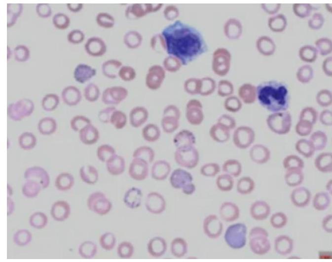
Which of the following diagnoses give the hematological picture as given below?

Which of the following statements is correct about Paroxysmal Nocturnal Hemoglobinuria (PNH)?
Explanation: ***1, 3 and 4*** - In **iron deficiency anemia**, **serum iron** levels are typically **less than 30 µg/dL** [1] (or 30 mg/100 mL), indicating a reduced iron supply. - The **percentage saturation** of transferrin with iron falls to **10% or less** [1] because there is insufficient iron to bind to the available transferrin. - **Serum ferritin**, which reflects iron stores, is significantly **reduced, usually below 30 ng/mL** (or 30 µg/mL) [1]. *1, 2 and 4* - While options 1 and 4 are correct, option 2 stating **total iron binding capacity (TIBC) less than 400 µg/mL** is incorrect. - In iron deficiency, the body attempts to increase iron absorption by producing more transferrin, leading to an **elevated TIBC** [1] (often >400 µg/dL). *1, 2 and 3* - Although options 1 and 3 are correct for iron deficiency anemia, option 2, which states **TIBC is less than 400 µg/mL**, is false. - **TIBC is elevated** in iron deficiency, reflecting an increased capacity for iron binding due to increased transferrin. *2, 3 and 4* - While options 3 and 4 are correct, option 2, suggesting **TIBC is less than 400 µg/mL**, is inaccurate. - **TIBC** is typically **increased** in iron deficiency anemia as the body tries to maximize any available iron.
Explanation: ***1, 2 and 4*** * **Iron deficiency** is indeed more common in **older age groups**, often due to decreased dietary intake, malabsorption, or chronic diseases [1], [3]. * It is also frequently observed in **females of reproductive age**, primarily due to physiological iron loss through menstruation [2]. * **Chronic blood loss** from various sources, such as gastrointestinal bleeding [1] or heavy menstruation [2], is a very common cause of iron deficiency [3]. *1, 2 and 3* * While iron deficiency is common in older age groups and females of reproductive age, the statement that it is *only* associated with heavy menstrual bleeding isn't comprehensive enough, as chronic blood loss is a broader and more common cause [1]. * Though heavy menstrual bleeding is a significant cause of iron deficiency in women, this option omits chronic blood loss as a more general and equally important cause. *2, 3 and 4* * This option incorrectly implies that iron deficiency is *not* more common in older age groups, which is a known demographic at risk [3]. * While **heavy menstrual bleeding** and **chronic blood loss** are major causes, excluding the increased prevalence in older populations makes this option incomplete. *1, 3 and 4* * This option inaccurately suggests that iron deficiency is *not* more common in females of reproductive age, which is a key demographic for iron deficiency due to menstrual blood loss [2]. * It correctly identifies chronic blood loss and heavy menstrual bleeding as causes but misses a significant demographic for prevalence.
Explanation: ***1 and 3 only*** - Haemophilia is a well-known **hereditary bleeding disorder**, primarily affecting males, caused by a deficiency in certain clotting factors [1]. - While haemophilia mainly affects males, females can be carriers and, in rare instances, experience mild to moderate symptoms, especially due to **extreme X-inactivation**. *2 and 3 only* - The statement that it has a higher incidence among Asians is **incorrect**; haemophilia incidence is generally consistent across ethnic groups, approximately 1 in 5,000 live male births worldwide [2]. - While females can be affected, their involvement is typically mild and occasional, making statement 3 correct. *1 and 2 only* - Haemophilia is indeed a **hereditary bleeding disorder** (statement 1 is correct) [1]. - However, the claim of higher incidence among Asians is **unsupported** by epidemiological data, rendering statement 2 incorrect. *1, 2 and 3* - While haemophilia is a **hereditary bleeding disorder** and can affect females, the assertion of a higher incidence among Asians is **false** [2]. - Its global incidence rates do not show a significant racial or ethnic predilection.
Explanation: ***Massive splenomegaly*** - Idiopathic Thrombocytopenic Purpura (ITP) typically involves only **mild to moderate splenomegaly**, if any, due to increased platelet destruction in the spleen [1]. **Massive splenomegaly** is not a characteristic feature. - **Massive splenomegaly** would suggest other conditions like chronic myeloid leukemia, myelofibrosis, or certain infectious diseases. *Cutaneous ecchymoses* - **Ecchymoses (bruising)** are a common symptom of ITP due to the low platelet count, leading to easier bleeding under the skin [2]. - These present as larger, purplish patches on the skin, indicating extravasated blood. *Thrombocytopenia* - **Thrombocytopenia** (low platelet count) is the hallmark and diagnostic criterion for ITP [1]. - The reduced number of platelets impairs the body's ability to form clots, leading to bleeding manifestations [2]. *Epistaxis* - **Epistaxis (nosebleeds)** is a common mucocutaneous bleeding symptom in ITP, reflecting the impaired primary hemostasis due to low platelet count [2]. - Bleeding from mucosal surfaces is typical in platelet disorders [2].
Explanation: ***Leukaemia*** - In leukaemia, especially **myeloid leukaemia**, immature white blood cells can accumulate in the **corpus cavernosa**, leading to stasis and **venous occlusion**. [1] - This cellular congestion prevents venous outflow from the penis, causing a prolonged and **painful erection (priapism)**. *Carcinoid tumour of appendix* - A carcinoid tumour of the appendix is typically associated with **carcinoid syndrome**, which involves symptoms like flushing and diarrhoea. - It does **not directly cause priapism**, as its mediators (e.g., serotonin) do not typically induce this specific local vascular event. *Testicular cancer* - Testicular cancer primarily manifests as a **painless lump in the testis** and can cause symptoms related to metastasis, but **priapism is not a typical direct presenting symptom**. - While some cancers can cause paraneoplastic syndromes, testicular cancer is not associated with priapism. *Penile cancer* - Penile cancer typically presents as a **lesion, ulcer, or mass on the penis**, often associated with pain, bleeding, or discharge. - While it affects the penis, it does **not typically cause prolonged erections (priapism)**, but rather local tissue destruction or obstruction.
Explanation: ***Parkinsonism*** - While stem cell research for Parkinson's disease is ongoing, current treatments using **umbilical cord blood stem cells** have not shown efficacy or routine use for this neurodegenerative disorder. - Parkinson's disease primarily affects **dopaminergic neurons** in the substantia nigra, which are not effectively replaced or regenerated by umbilical cord blood stem cells in a therapeutic context. *Leukemia* - **Umbilical cord blood stem cells** are a well-established source of **hematopoietic stem cells** used in transplantation to treat various forms of leukemia. - These stem cells can engraft in the bone marrow and differentiate into healthy blood cells, replacing cancerous cells after chemotherapy or radiation. *Osteoporosis* - Research is exploring the use of **mesenchymal stem cells** from umbilical cord blood for bone regeneration and treatment of osteoporosis. - These stem cells have the potential to differentiate into **osteoblasts**, which are essential for bone formation and repair. *Diabetes* - Experimental treatments and clinical trials are investigating the use of **umbilical cord blood stem cells** to treat Type 1 Diabetes by modulating the immune system and potentially regenerating insulin-producing beta cells. - The immunomodulatory properties of these stem cells may help in preventing the autoimmune destruction of **pancreatic beta cells**.
Explanation: ***2 and 3*** - Hemophilia A is caused by a deficiency of **Factor VIII**, and Hemophilia B is caused by a deficiency of **Factor IX** [1]. These are distinct but clinically similar genetic disorders of the coagulation cascade. - Both hemophilia A and B are **X-linked recessive disorders**, meaning they predominantly affect males and are transmitted by asymptomatic female carriers [2]. *1, 2 and 4* - While both hemophilia A and B result in similar bleeding phenotypes, they are due to deficiencies in different **coagulation factors** (Factor VIII and Factor IX, respectively), making them distinct diseases, not variants of the same process [2]. - While hemophilia mainly affects males, a female can be affected if she inherits two affected X chromosomes (one from each parent) or has **Turner syndrome** (XO) with an affected X chromosome, but this is extremely rare. *2 only* - This option correctly identifies the specific factor deficiencies for Hemophilia A (**Factor VIII**) and Hemophilia B (**Factor IX**) [1]. - However, it omits the crucial aspect of their **sex-linked inheritance** and transmission by asymptomatic females, which is a fundamental characteristic of both conditions [2]. *3 only* - This option correctly states that both hemophilia A and B are **sex-linked characteristics** and are transmitted by **asymptomatic females** [2]. - However, it fails to mention the specific deficient factors (Factor VIII and Factor IX), which is key to understanding the underlying pathology of these two distinct disorders.
Explanation: ***4°C*** - Whole blood is typically stored at **1 to 6°C**, with **4°C** being the optimal compromise to preserve red blood cell viability and minimize bacterial growth [1]. - This temperature range allows for a standard storage duration of **21 to 42 days**, depending on the anticoagulant-preservative solution used [1]. *-4°C* - Temperatures below freezing point (**0°C**) would cause **hemolysis** due to ice crystal formation within the red blood cells, making the blood unsuitable for transfusion. - While frozen storage is used for specific blood components (e.g., cryopreserved red blood cells or plasma), it requires different protocols and cryoprotectants. *8°C* - Storing whole blood at **8°C** is above the recommended range and would significantly decrease the storage time due to increased metabolic activity and **bacterial proliferation**. - This temperature also leads to a faster decline in **red blood cell viability** and function. *0°C* - Storing whole blood at **0°C** is at the freezing point of water and close to the freezing point of blood, which can lead to initial **ice crystal formation** and subsequent hemolysis. - Although it's within the recommended 1-6°C range, maintaining exactly 0°C without fluctuations into freezing territory is challenging and poses a risk to blood quality [1].
Explanation: ***Saturnism*** - The image displays **basophilic stippling** in red blood cells, which is a classic hematological finding in **lead poisoning** (saturnism). - Lead inhibits enzymes involved in **heme synthesis**, leading to the accumulation of ribosomal RNA aggregates detected as basophilic stippling. *Arsenic poisoning* - Arsenic poisoning can cause various hematological abnormalities, including **anemia** and **pancytopenia**, but **basophilic stippling** is not a characteristic feature. - Its mechanism of toxicity involves inhibiting enzyme function and cellular respiration, distinct from lead's effect on heme synthesis. *Chronic iron toxicity* - Chronic iron toxicity typically leads to **hemochromatosis**, with iron deposition in various organs, and can cause **liver damage** and **cardiomyopathy**. - It does not primarily manifest with **basophilic stippling** in red blood cells. *Minamata disease* - Minamata disease is a severe neurological syndrome caused by **mercury poisoning**, particularly **methylmercury**. - It primarily affects the **nervous system**, causing symptoms like ataxia, sensory disturbances, and tremors, and does not typically present with **basophilic stippling**.
Explanation: ***Flow cytometry is the best investigation*** - **Flow cytometry** is the gold standard for diagnosing PNH as it directly detects the absence of **GPI-anchored proteins (CD55 and CD59)** on the surface of red blood cells, granulocytes, and monocytes. - This method is highly sensitive and specific, allowing for the precise quantification of PNH clones even when present in small numbers. *Sucrose lysis test is used for the confirmation* - The **sucrose lysis test** (or sugar-water test) was an older screening test for PNH, but it is **not specific or sensitive enough** for confirmation. - It works by inducing complement activation on PNH cells in low ionic strength sucrose solution [1], but it has a high rate of false positives and negatives. *The most common cause of death is heart failure* - The most common cause of death in patients with PNH is **thrombosis**, particularly in unusual sites such as hepatic, cerebral, or abdominal veins, due to the prothrombotic state caused by the disease. - While heart complications can occur, they are not the leading cause of mortality. *It is an inherited disorder* - PNH is an **acquired clonal disorder** of hematopoietic stem cells, not an inherited genetic disorder [1]. - It results from a somatic mutation in the **PIG-A gene** in a hematopoietic stem cell, leading to the deficiency of GPI-anchored proteins.
Anemia Evaluation and Management
Practice Questions
Hemoglobinopathies
Practice Questions
Thalassemias
Practice Questions
Platelet Disorders
Practice Questions
Coagulation Disorders
Practice Questions
Thrombotic Disorders
Practice Questions
Leukemias
Practice Questions
Lymphomas
Practice Questions
Multiple Myeloma and Plasma Cell Disorders
Practice Questions
Myeloproliferative Neoplasms
Practice Questions
Transfusion Medicine
Practice Questions
Hematopoietic Stem Cell Transplantation
Practice Questions
Get full access to all questions, explanations, and performance tracking.
Start For Free