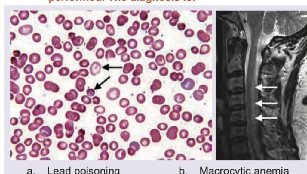
A 45-year-old person has complaints of paresthesia in hand and feet with progressive spastic weakness. On examination, absent ankle jerk with Babinski sign is noted. Peripheral smear and MRI spine were performed. The diagnosis is:

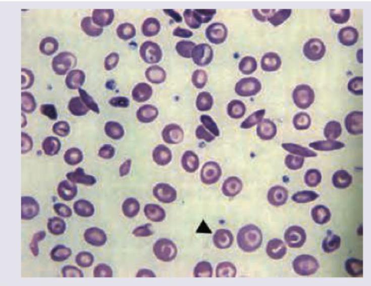
A 16-year-old female with pallor and hepatosplenomegaly presents to your clinic. Peripheral smear shows:

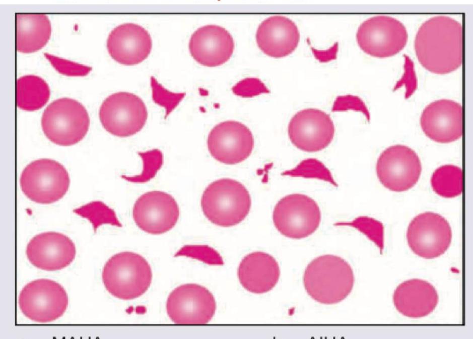
Which is shown in the peripheral smear of patient with metallic heart prosthesis?

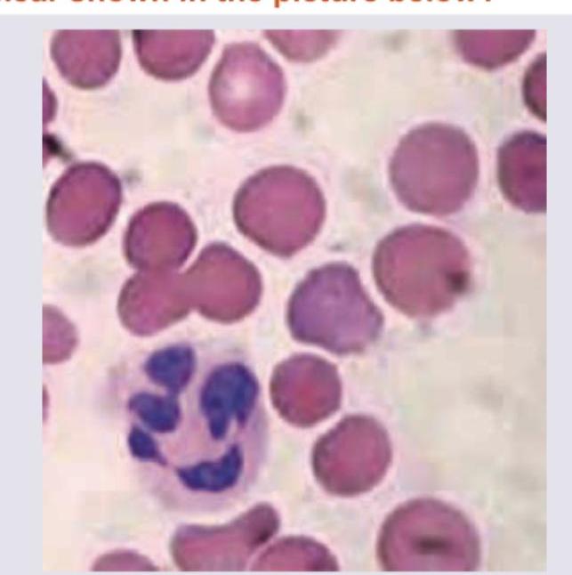
Which of the following is seen with the peripheral smear shown in the picture below?

Which of the following treatments are recommended for a pregnant woman suffering from sickle cell disease ? I. Folic acid 1 mg daily II. Azathioprine III. Penicillin prophylaxis IV. Thromboprophylaxis with low molecular weight heparin Select the correct answer using the code given below :
Which of the following are secondary iron overload conditions? I. Transfusion related iron load II. Thalassemia III. Hepatitis C associated liver disease Select the correct answer using the code given below :
With regard to management of idiopathic thrombocytopenic purpura, consider the following statements : I. All patients with ITP with platelet count less than 100 x 10^9 / L should receive high dose of glucocorticoids II. For patient with spontaneous bleeding, 40 mg of dexamethasone daily is indicated for 4 days III. Intravenous immunoglobulin can raise the platelet count IV. Life threatening bleeding should be treated with platelet transfusion Which of the statements given above are correct?
First line therapy in chronic phase of chronic myeloid leukemia is
Which one of the following causes low-volume erythrocytosis?
A 35 year female presents with easy fatigability. Investigations show that Hb is 6 g %; Red cell morphology is normocytic normochromic; and reticulocyte production index is 5.5 . Which one of the following conditions favours this abnormality?
Explanation: ***Macrocytic anemia*** - The peripheral smear shows **macrocytic red blood cells** and possibly **hypersegmented neutrophils** (though not clearly visible in this image alone, the arrows *could* be pointing to basophilic stippling often seen in lead poisoning or other erythrocyte inclusions related to dyserythropoiesis in megaloblastic anemia). The MRI of the spine shows **posterior column degeneration** (white arrows indicating increased signal intensity), which is characteristic of **subacute combined degeneration** of the spinal cord seen in severe **vitamin B12 deficiency** (a common cause of macrocytic anemia). - The neurological symptoms of paresthesia, progressive spastic weakness, absent ankle jerk, and a positive Babinski sign are all consistent with the combined peripheral neuropathy and myelopathy (damage to posterior and lateral columns) associated with **vitamin B12 deficiency**. *Lead poisoning* - While lead poisoning can cause neurological symptoms and anemia, the anemia is typically **microcytic hypochromic** or normocytic with prominent **basophilic stippling** on peripheral smear. - Lead poisoning does not typically lead to the distinct pattern of **spinal cord degeneration** seen on the MRI. *Miller Fisher syndrome* - This is a rare variant of Guillain-Barré syndrome characterized by the triad of **ataxia**, **arreflexia**, and **ophthalmoplegia**. - It primarily affects peripheral nerves and cranial nerves, not typically causing progressive spastic weakness with pyramidal signs like **Babinski sign** or central demyelination seen on MRI. *Devic's disease* - Also known as neuromyelitis optica, Devic's disease is an inflammatory demyelinating condition that primarily affects the **optic nerves** and **spinal cord**. - While it causes myelopathy (and thus spasticity and Babinski sign) and can have MRI findings in the spinal cord, it is not associated with **macrocytic anemia** or abnormal peripheral blood smears, and features like absent ankle jerk (suggesting peripheral neuropathy) are not typical primary findings.
Explanation: ***Thalassemia intermedia*** - The peripheral smear shows significant **anisopoikilocytosis** with numerous **target cells** (indicated by the arrowhead), teardrop cells, and some irregularly contracted cells, which are characteristic of thalassemia. - The patient's presentation with **pallor** and **hepatosplenomegaly** (due to extramedullary hematopoiesis and increased red cell destruction) in a 16-year-old female points towards a chronic hemolytic anemia like thalassemia intermedia. *Iron-deficiency anemia* - While it causes **microcytic hypochromic anemia**, the peripheral smear would typically show predominantly small, pale red cells and not the extensive target cells, teardrop cells, or significant anisopoikilocytosis seen in the image. - **Hepatosplenomegaly** is not a common finding in uncomplicated iron-deficiency anemia. *Autoimmune hemolytic anemia* - This condition typically presents with **spherocytes** (small, dense red cells lacking central pallor) due to immune-mediated destruction, and sometimes **agglutination**, which are not the predominant findings in this smear. - While it can cause anemia and splenomegaly, the distinct red cell morphology in the image is not typical for AIHA. *Megaloblastic anemia* - The classic peripheral smear findings for megaloblastic anemia are **macrocytic red blood cells** and **hypersegmented neutrophils**, neither of which are observed in this image. - The presence of target cells and other poikilocytes seen here excludes megaloblastic anemia.
Explanation: ***MAHA*** - Metallic heart valve prostheses can cause **Microangiopathic Hemolytic Anemia (MAHA)** due to mechanical shearing of red blood cells as they pass through the turbulent flow around the valve. - The peripheral smear in MAHA typically shows **schistocytes** (fragmented red blood cells), which are represented by the irregular, fragmented pink structures in the image, mixed with intact red blood cells. *AIHA* - **Autoimmune Hemolytic Anemia (AIHA)** is characterized by immune-mediated red blood cell destruction, often showing **spherocytes** on the peripheral smear, which are not depicted here. - The mechanism involves antibodies attacking red blood cells, rather than mechanical destruction. *Echinocytes* - **Echinocytes (burr cells)** are red blood cells with uniformly spaced, short, blunt projections on their surface, often seen in uremia or artifactually. - The fragmented cells shown in the image are distinctly different from echinocytes. *Dimorphic anemia* - **Dimorphic anemia** is a condition where two distinct populations of red blood cells (e.g., microcytic hypochromic and macrocytic) are present, typically due to combined deficiencies like iron and folate/B12. - The image mainly shows fragmented red blood cells and normal red blood cells, not two distinct populations based on size and hemoglobin content.
Explanation: ***Beefy tongue*** - The peripheral smear shows **hypersegmented neutrophils** and **macrocytic red blood cells**, which are characteristic findings in **megaloblastic anemia**. - **Beefy tongue** (glossitis) is a classic oral manifestation of **vitamin B12 deficiency**, a common cause of megaloblastic anemia. *Magenta tongue* - **Magenta tongue** is typically associated with **riboflavin (vitamin B2) deficiency**, not megaloblastic anemia. - While nutritional deficiencies can coexist, this specific tongue presentation is not directly linked to the red blood cell morphology shown. *Geographic tongue* - **Geographic tongue** (benign migratory glossitis) is a common inflammatory condition of the tongue characterized by irregular, red patches that resemble maps. - It is generally considered benign and not directly associated with the hematological findings seen in the image. *Black hairy tongue* - **Black hairy tongue** is a harmless condition where the papillae on the tongue become elongated and discolored, often due to poor oral hygiene, smoking, or certain medications. - This condition is not related to the hematological abnormalities depicted in the peripheral smear.
Explanation: ***I, III and IV*** - **Folic acid 5 mg daily** (not 1mg) is essential in pregnancy, especially for women with **sickle cell disease**, to prevent **megaloblastic anemia** due to increased red cell turnover [1]. - **Penicillin prophylaxis** is crucial to prevent **Bacterial infections** as patients with sickle cell disease are at increased risk of infection, especially from encapsulated organisms, due to **functional asplenia** [1]. - **Thromboprophylaxis with low molecular weight heparin** is recommended because pregnancy in sickle cell disease significantly increases the risk of **venous thromboembolism** [1]. *I, II and IV* - **Azathioprine** is an immunosuppressant typically used for autoimmune conditions or organ transplantation, and it is **not a standard treatment** for managing sickle cell disease itself during pregnancy. - While folic acid and thromboprophylaxis are indicated, the inclusion of azathioprine makes this option incorrect. *I, II and III* - This option incorrectly includes **azathioprine**, an immunosuppressant not indicated for routine sickle cell management in pregnancy. - It also omits crucial **thromboprophylaxis**, which is vital given the increased risk of blood clots. *II, III and IV* - This option correctly includes **penicillin prophylaxis** and **thromboprophylaxis** but **incorrectly omits folic acid**, which is a cornerstone of daily management for all pregnant women with sickle cell disease. - It also incorrectly includes **azathioprine**, which is not a standard treatment.
Explanation: ***I, II and III*** - All three listed conditions—**transfusion-related iron load**, **thalassemia**, and **Hepatitis C associated liver disease**—are well-recognized causes of secondary iron overload. - Secondary iron overload occurs due to external factors or underlying diseases that cause increased iron absorption or repeated administration of iron. [1] *I and II only* - This option is incomplete as **Hepatitis C associated liver disease** can also lead to secondary iron overload due to impaired iron metabolism and chronic inflammation. - It dismisses a known cause of secondary iron accumulation. *I and III only* - This option is incomplete because **thalassemia**, particularly **transfusion-dependent thalassemia**, is a classic example of secondary iron overload due to frequent blood transfusions and ineffective erythropoiesis. - It overlooks a major cause of transfusion-related iron accumulation. [1] *II and III only* - This option is incomplete as **transfusion-related iron load** is a direct and common cause of secondary iron overload, especially in patients requiring regular blood transfusions for conditions like anemia. [1] - It ignores the most direct mechanism of iron accumulation in many chronic diseases.
Explanation: ***II, III and IV*** - Statement II is correct because **40 mg of dexamethasone daily for 4 days** is a common regimen for newly diagnosed ITP with a platelet count <30 x 10^9/L or with bleeding, aiming for rapid platelet increase. - Statement III is correct as **Intravenous Immunoglobulin (IVIG)** works by blocking Fc receptors on macrophages, reducing platelet destruction and rapidly raising platelet counts, especially in cases of active bleeding or before urgent procedures. - Statement IV is correct because in **life-threatening bleeding** associated with ITP, platelet transfusions are indicated to provide immediate hemostasis, often given alongside other therapies like IVIG or high-dose steroids. *I, II and IV* - Statement I is incorrect; not all ITP patients with platelet counts <100 x 10^9/L require high-dose glucocorticoids [1]. Treatment decisions are based on **platelet count, bleeding symptoms**, and patient risk factors, with many asymptomatic patients with counts between 30-100 x 10^9/L being observed. - While statements II and IV are correct, the inclusion of statement I makes this option incorrect. *I, II and III* - Statement I is incorrect for the reasons mentioned above; not all ITP patients with platelet counts <100 x 10^9/L need high-dose glucocorticoids. - Although statements II and III are correct, the presence of the incorrect statement I renders this option invalid. *I, III and IV* - Statement I is incorrect because immediate high-dose glucocorticoid treatment is not indicated for all ITP patients with platelet counts below 100 x 10^9/L; it's reserved for those with **significant bleeding or very low counts** (<30 x 10^9/L) [1]. - Statements III and IV are correct, but the inaccuracy of statement I makes this choice incorrect.
Explanation: ***Imatinib*** - **Imatinib** is a **tyrosine kinase inhibitor (TKI)** that specifically targets the **BCR-ABL fusion protein**, which is the hallmark of **chronic myeloid leukemia (CML)** [1][2]. - It is highly effective in inducing **hematologic and cytogenetic remissions** in the chronic phase of CML and has significantly improved prognosis [2]. *Thalidomide* - **Thalidomide** is an immunomodulatory drug primarily used in **multiple myeloma** and as a teratogen. - It does not target the **BCR-ABL fusion protein** and is not indicated for CML. *Rituximab* - **Rituximab** is a **monoclonal antibody** that targets the **CD20 antigen** found on B-lymphocytes. - It is used in the treatment of **B-cell non-Hodgkin lymphoma** and **chronic lymphocytic leukemia**, not CML. *Chlorambucil* - **Chlorambucil** is an **alkylating agent**, a type of chemotherapy drug. - While historically used in some hematologic malignancies, it has been largely superseded by targeted therapies like TKIs in CML due to its non-specific action and greater toxicity [2].
Explanation: ***Gaisbock's syndrome*** - **Gaisbock's syndrome**, also known as **stress erythrocytosis** or **relative polycythemia**, is characterized by a high hematocrit due to reduced plasma volume rather than an absolute increase in red blood cell (RBC) mass. - It is often associated with **hypertension**, **obesity**, and **stress**, predominantly affecting middle-aged men with a normal total erythrocyte mass [1]. *High altitude* - Living at **high altitudes** can cause **secondary erythrocytosis** due to chronic hypoxia, leading to increased erythropoietin production and an absolute increase in red blood cell mass [2]. - This is a **true erythrocytosis**, where both red blood cell count and total blood volume are elevated. *Polycythemia Rubra Vera* - **Polycythemia vera** is a **myeloproliferative neoplasm** causing an absolute increase in the red blood cell mass, resulting from uncontrolled production by the bone marrow. - It is characterized by a **JAK2V617F mutation** and leads to increased total blood volume, not reduced plasma volume [1]. *Exogenous testosterone therapy* - **Exogenous testosterone therapy** can stimulate erythropoiesis, leading to an **increase in red blood cell mass** and hematocrit, which is a form of **secondary erythrocytosis**. - This effect is mediated by increased erythropoietin production and is an absolute increase in RBCs, not a low-volume condition.
Explanation: ***Intravascular hemolysis*** - **Normocytic, normochromic anemia** with a **high reticulocyte production index** (RPI > 2-3) is characteristic of hemolytic anemias, indicating the bone marrow is effectively responding to increased red cell destruction [1]. - **Easy fatigability** is a common symptom of severe anemia, and the low Hb of 6g% confirms significant red blood cell loss, consistent with active hemolysis. *Folate deficiency* - This typically causes **macrocytic anemia** with **megaloblastic red cell morphology**, which contradicts the given normocytic finding [2]. - While it can lead to anemia and fatigue, the **reticulocyte index would be low**, indicating impaired red blood cell production, not increased destruction. *Sideroblastic anemia* - This anemia is usually **microcytic or dimorphic** and can be normocytic, but it is characterized by **ring sideroblasts** in the bone marrow, reflecting impaired heme synthesis. - The **reticulocyte production index is typically low or inappropriately normal**, as the primary issue is ineffective erythropoiesis rather than rapid peripheral destruction. *Iron deficiency anemia* - This is classically a **microcytic, hypochromic anemia**, meaning red blood cells are smaller and paler than normal [3]. - The **reticulocyte production index would be low or normal**, as the bone marrow's ability to produce new red blood cells is limited by the lack of iron.
Anemia Evaluation and Management
Practice Questions
Hemoglobinopathies
Practice Questions
Thalassemias
Practice Questions
Platelet Disorders
Practice Questions
Coagulation Disorders
Practice Questions
Thrombotic Disorders
Practice Questions
Leukemias
Practice Questions
Lymphomas
Practice Questions
Multiple Myeloma and Plasma Cell Disorders
Practice Questions
Myeloproliferative Neoplasms
Practice Questions
Transfusion Medicine
Practice Questions
Hematopoietic Stem Cell Transplantation
Practice Questions
Get full access to all questions, explanations, and performance tracking.
Start For Free