Which of the following is not included in the Gaisböck syndrome?
A 38-year-old female presents with fatigue, weakness, and pallor for 3 months. She has a history of menorrhagia. Laboratory findings show hemoglobin 8.5 g/dL, MCV 68 fL, MCH 22 pg, serum ferritin 8 ng/mL, serum iron 30 μg/dL, TIBC 450 μg/dL, and peripheral smear shows microcytic hypochromic RBCs with pencil cells. Bone marrow examination shows absent iron stores. What is the most appropriate next step in management?
A 32-year-old woman presents with easy bruising and petechiae. Her platelet count is 18,000 / mm³. Hemoglobin and total leukocyte count are normal. She recently recovered from a mild upper respiratory tract infection. Peripheral smear shows no abnormal cells. What is the most appropriate next step in management?
A 16-year-old boy presents with jaundice and splenomegaly. His father had a similar illness during adolescence. MCHC : high? What is the most likely diagnosis?
Which of the following is the most common inherited bleeding disorder?
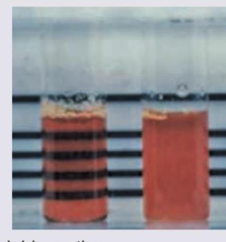
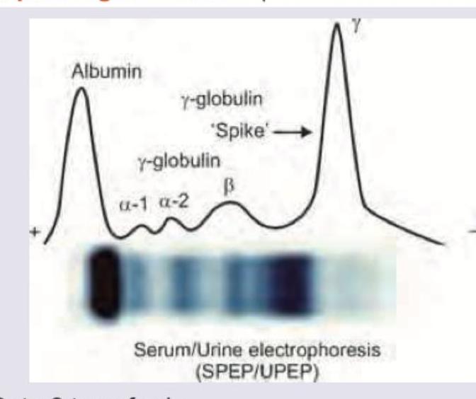
The following test is performed for which condition?

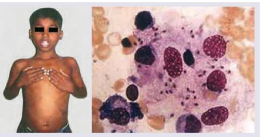
An 8-year-old child of migrant construction worker presents with fever for last 2 weeks with progressive pallor. Complete blood counts reveal pancytopenia, malaria work up is negative, Total serum protein is 7 gm% with Serum Globulin of 4.5 gm%, serum albumin 3.5 mg%. Bone marrow study shows?

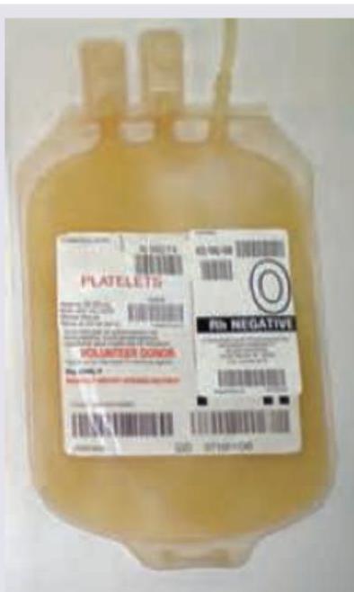
All are true about the blood component shown below except?

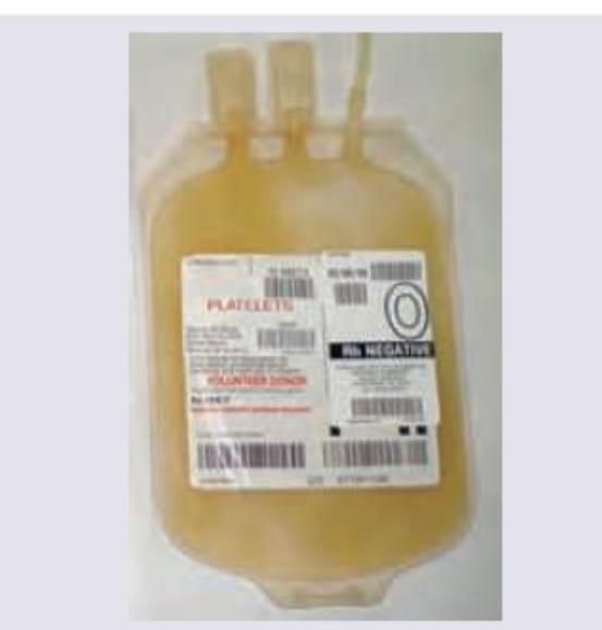
What is the threshold for prophylactic administration of the following blood product?

Which of the following is a prognostic marker for the patient whose urinary and serum electrophoresis report is given below?

Explanation: Hypotension- Gaisböck syndrome, often called stress polycythemia or relative polycythemia, is strongly associated with hypertension due to the hemoconcentration and reduced plasma volume [1].- Hypotension is not a feature of this syndrome, which typically presents in middle-aged men with cardiovascular risk factors.*Erythrocytosis*- Relative erythrocytosis (or pseudopolycythemia) is the defining characteristic of Gaisböck syndrome [1].- This increase in hematocrit is due to a decrease in plasma volume (hemoconcentration) rather than an absolute increase in red cell mass.*Normal leukocyte counts*- Maintaining normal white blood cell (WBC) counts is typical in Gaisböck syndrome [1].- This feature helps exclude Polycythemia Vera, where both leukocytes and platelets are often elevated along with the absolute erythrocytosis.*Obesity*- Obesity is a common and significant component of the syndrome, along with other metabolic risk factors like hyperlipidemia.- The patient group is often described as overweight, anxious, and highly prone to cardiovascular events.
Explanation: The laboratory findings (low Hb, low MCV, low MCH, significantly low **serum ferritin**, high **TIBC**) are classic for severe **Iron Deficiency Anemia (IDA)** [1]. The most appropriate first-line treatment for uncomplicated IDA, especially in a patient with chronic blood loss (menorrhagia), is high-dose **oral ferrous salts** (e.g., ferrous sulfate) [1]. Menorrhagia is a common cause of anaemia in pre-menopausal females [2]. Elevated hepcidin in anemia of inflammation can make it refractory to iron supplementation, making the diagnosis of true IDA important [3]. Blood transfusion is indicated for patients with anemia causing **hemodynamic instability**, impending **cardiac decompensation**, or extremely low Hgb levels (often <7 g/dL) [4].
Explanation: ***Oral corticosteroids*** - This is the **first-line treatment** for newly diagnosed Immune Thrombocytopenic Purpura (ITP), especially when the platelet count is severely low (typically <30,000/mm³) and the patient exhibits bleeding symptoms like **petechiae** and easy bruising [1]. - Corticosteroids reduce the clearance of antibody-coated platelets by the **reticuloendothelial system** (primarily splenic macrophages) and suppress autoantibody production. ***Platelet transfusion*** - Platelet transfusions are generally **contraindicated** in ITP because the transfused platelets are rapidly destroyed by the same autoantibodies that target the patient's native platelets [1]. - They are reserved only for **life-threatening hemorrhage** (e.g., intracranial bleeding) or for pre-operative stabilization, usually given concurrently with high-dose IVIG or corticosteroids. ***Bone marrow biopsy*** - A **bone marrow biopsy** is usually not required for a young patient with classic ITP presentation (isolated thrombocytopenia, normal Hb/TLC, recent infection, and normal peripheral smear). - It is typically mandatory only if the patient is older (e.g., >60), has unexplained cytopenias in other lineages, or is **refractory** to first-line treatment. ***Splenectomy*** - Splenectomy is a **second-line treatment** option, typically reserved for patients with chronic ITP who fail to respond adequately to medical therapy (corticosteroids and IVIG) after 6 to 12 months [2]. - Although the spleen is the primary site of autoantibody production and platelet destruction, it is not the **most appropriate immediate next step** in initial management [2].
Explanation: ***Hereditary spherocytosis*** - The presentation of **jaundice**, **splenomegaly**, strong **family history** (autosomal dominant pattern), and an elevated **MCHC** is classic for Hereditary Spherocytosis. [1] - Elevated MCHC is a key diagnostic feature due to mild cellular dehydration in the characteristic **spherocytes**. [1] *IDA* - **Iron Deficiency Anemia (IDA)** is typically microcytic and hypochromic, leading to a **low MCHC**, not high. - Symptoms of IDA include fatigue and pallor, generally without marked jaundice or splenomegaly unless severe. *Thalassemia major* - **Thalassemia major** presents with severe microcytic, hypochromic anemia (low MCHC) usually evident in early infancy, requiring regular transfusions. - While it causes jaundice and hepatosplenomegaly, it is characterized by severe bone changes and a very low MCHC. *AIHA* - **Autoimmune Hemolytic Anemia (AIHA)** typically presents acutely or subacutely and often responds to immunosuppression (e.g., steroids). - Unlike Hereditary Spherocytosis, AIHA generally does not present with a positive **family history** and the primary lab finding is a positive **Direct Antiglobulin Test (DAT)**, not specifically high MCHC.
Explanation: ***Von Willebrand Disease*** - VWD is the most common inherited bleeding disorder, with a prevalence estimated to be as high as 1% of the population, often showing an **autosomal dominant** inheritance pattern. - It results from a quantitative or qualitative defect in **von Willebrand factor (vWF)**, which impairs platelet adhesion and stabilizes **Factor VIII** [1][2]. *Disseminated Intravascular Coagulation* - DIC is an **acquired** life-threatening condition caused by systemic activation of coagulation, usually triggered by underlying severe diseases like **sepsis** or trauma, not inheritance. - It is characterized by simultaneous widespread thrombosis and severe bleeding due to the consumption of clotting factors and platelets. *Factor V Deficiency* - Factor V deficiency (Parahemophilia) is an extremely rare inherited bleeding disorder, transmitted in an **autosomal recessive** manner. - While inherited, its overall prevalence is substantially lower compared to VWD, making it not the most common. *Hemophilia* - Hemophilia (A and B) is the most common severe inherited bleeding disorder, known for causing deep tissue bleeding (e.g., **hemarthroses**). - It is an **X-linked recessive** condition, meaning it is less prevalent overall in the population compared to the high frequency of mild, often undiagnosed, VWD cases.
Explanation: ***Paroxysmal nocturnal haemoglobinuria*** - The image depicts the **Ham test**, also known as the acid serum lysis test, which is historically used to diagnose **paroxysmal nocturnal hemoglobinuria (PNH)**. - In PNH, red blood cells are abnormally susceptible to complement-mediated lysis in acidified serum, leading to **hemolysis** as seen in the test tube with red fluid. *Hemoglobinopathy* - Hemoglobinopathies are characterized by **abnormal hemoglobin structures** or synthesis, such as sickle cell anemia or thalassemia. - While they can cause hemolytic anemia, their diagnosis involves **hemoglobin electrophoresis** or genetic testing, not the Ham test. *Alkaptonuria* - Alkaptonuria is a rare genetic disorder of metabolism affecting the breakdown of **tyrosine and phenylalanine**, leading to accumulation of homogentisic acid. - It results in **dark urine** upon standing and pigmentation of connective tissues (ochronosis), and is not diagnosed by tests for hemolysis. *Paroxysmal cold haemoglobinuria* - This condition is characterized by **hemolysis following cold exposure**, mediated by anti-P cold agglutinin antibodies. - Diagnosis involves the **Donath-Landsteiner test**, which demonstrates hemolysis at cold temperatures and warming, rather than the Ham test.
Explanation: ***L.D bodies*** - The image provided shows **macrophages** laden with numerous **Leishman-Donovan bodies**, which are intracellular amastigotes of *Leishmania* parasites. This bone marrow finding, in conjunction with **pancytopenia** and **fever with progressive pallor** in a child from a region where migrant workers live (suggesting possible exposure to endemic diseases), points strongly to **visceral leishmaniasis (kala-azar)**. - The elevated serum globulin and positive total serum protein differentiate from conditions like aplastic anemia. *Aplastic crisis* - An aplastic crisis results in **pancytopenia** but is characterized by a **hypocellular bone marrow** with a severe reduction in hematopoietic precursors. - It would not show the presence of **L.D. bodies** or hypergammaglobulinemia. *Myelofibrosis* - Myelofibrosis is characterized by **fibrotic bone marrow** and usually presents with cytopenias, but also often with **splenomegaly** and a leukoerythroblastic peripheral smear. - The bone marrow would show *fibrosis* and not parasitic inclusions. *Myeloid metaplasia* - Myeloid metaplasia (extramedullary hematopoiesis) is the presence of hematopoietic tissue outside the bone marrow, typically in the spleen or liver, and occurs in response to severe bone marrow dysfunction (e.g., myelofibrosis). - While it can be associated with pancytopenia, the primary bone marrow pathology here is parasitic infection, not a compensatory shift in hematopoiesis.
Explanation: ***Random donor platelets are produced by apheresis*** - **Random donor platelets** are typically prepared from **whole blood donations** by centrifugation, not apheresis. - **Apheresis** is a process used to collect specific blood components, such as platelets, from a single donor, resulting in a single-donor apheresis platelet unit, not random donor platelets. *Infusion must be completed within 30 minutes* - The recommended infusion time for platelets is generally **30-60 minutes**. - Rapid infusion (under 30 minutes) is usually not recommended and could potentially lead to adverse reactions or compromise platelet viability. *Use of leukocyte filters helps in reducing allosensitization* - **Leukocyte reduction** significantly reduces the number of white blood cells in platelet products. - This helps to decrease the risk of **febrile non-hemolytic transfusion reactions** and **alloimmunization** (allosensitization) to HLA antigens. *Two units of transfused platelets per square meter body surface area are anticipated to increase the platelet count by approximately 10,000/μL* - This statement accurately describes the expected **platelet increment** from standard random donor platelet transfusions. - The increment can vary based on factors such as patient condition and consumption, but this is a general guideline for adequate response.
Explanation: ***<10,000*** - The image clearly shows a bag labeled "PLATELETS." Prophylactic platelet transfusions are generally recommended when the **platelet count drops below 10,000/µL** in hemodynamically stable patients without additional risk factors for bleeding. - This threshold helps prevent **spontaneous hemorrhage**, especially in critically ill patients, as severe thrombocytopenia significantly increases the risk of serious bleeding events like intracranial hemorrhage. *<5,00* - A platelet count of **less than 5,000/µL** is an extremely low threshold, indicating a very high risk of spontaneous bleeding. - While transfusion would certainly be indicated at this level, the typical prophylactic threshold to prevent bleeding in stable patients is higher, at 10,000/µL. *<20,000* - A platelet count of **less than 20,000/µL** is a common threshold for prophylactic platelet transfusion when there are additional risk factors for bleeding, such as fever, sepsis, or certain medications. - However, for general prophylactic transfusion in *stable* patients without these additional risk factors, the lower threshold of <10,000/µL is usually applied. *<50,000* - A platelet count of **less than 50,000/µL** is typically the threshold for prophylactic platelet transfusion in patients who are undergoing (or about to undergo) an **invasive procedure** or surgery where hemostasis is critical. - It is not the general prophylactic threshold for preventing spontaneous bleeding in stable, non-surgical patients.
Explanation: ***Beta 2 microglobulin*** - The serum/urine electrophoresis shows a **monoclonal gamma spike**, which is characteristic of a **monoclonal gammopathy**, such as **multiple myeloma**. - **Beta 2 microglobulin** is a prognostic marker for multiple myeloma, reflecting **tumor burden** and **renal function**. *Beta 2 transferrin* - **Beta 2 transferrin** is a unique isoform of transferrin found in **cerebrospinal fluid (CSF)** and is used to detect CSF leaks. - It has no known role as a prognostic marker in **monoclonal gammopathies**. *Beta 2 glycoprotein* - **Beta 2 glycoprotein I** is involved in the coagulation cascade and is primarily associated with **antiphospholipid syndrome**. - It is not a prognostic marker for **monoclonal gammopathies**. *Beta 2 microalbumin* - The term **microalbumin** refers to a small amount of albumin in the urine, indicating **early kidney damage**. - While kidney involvement can occur in multiple myeloma, **beta 2 microalbumin** itself is not the primary prognostic marker in this context.
Anemia Evaluation and Management
Practice Questions
Hemoglobinopathies
Practice Questions
Thalassemias
Practice Questions
Platelet Disorders
Practice Questions
Coagulation Disorders
Practice Questions
Thrombotic Disorders
Practice Questions
Leukemias
Practice Questions
Lymphomas
Practice Questions
Multiple Myeloma and Plasma Cell Disorders
Practice Questions
Myeloproliferative Neoplasms
Practice Questions
Transfusion Medicine
Practice Questions
Hematopoietic Stem Cell Transplantation
Practice Questions
Get full access to all questions, explanations, and performance tracking.
Start For Free