Which of the following does not cause intravascular hemolysis?
Mentzer index more than 13 suggests a diagnosis of
Which of the following statements about Hematochromatosis is true?
What is used for exchange blood transfusion?
Sickle cell anemia leads to resistance towards?
Female on carbimazole therapy presents with sudden fever, rigors and sore throat. Which is the investigation of choice for this patient?
A young patient presents with a large retroperitoneal hemorrhage and a history of intermittent knee swelling after strenuous exercise. There is no history of mucosal bleeding. Which of the following clotting factors is primarily deficient in Hemophilia A?
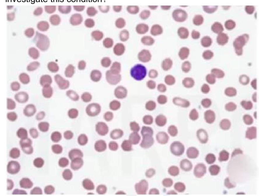
A child presents with intermittent jaundice and splenomegaly. There is a history of similar complaints in the elder brother. Peripheral smear shows the following finding. What is the most appropriate investigation for this condition?

A female patient presented with fatigue and a history of piles. Routine complete blood count analysis showed hemoglobin of 9 g/dL, MCV 60fL, and RBC count of 5.2 million. A peripheral smear is provided. Which of the following is the next best investigation after the smear for this patient?

In which condition are solitary lytic lesions typically seen?
Explanation: ***Hereditary spherocytosis*** - It primarily causes **extravascular hemolysis** due to spleen sequestration of **abnormal erythrocytes** [2]. - Not typically associated with **intravascular hemolysis**, making it the correct choice here. *Mismatched blood transfusion* - Causes acute **intravascular hemolysis** due to **antibody-mediated destruction** of foreign red blood cells [1]. - Results in **hemoglobinuria**, high bilirubin levels, and potential **renal failure**. *Paroxysmal cold hemoglobinuria* - Induces **intravascular hemolysis** triggered by cold temperatures and **anti-P antibodies** [3]. - Characterized by **acute hemoglobinuria** upon rewarming, complicating the clinical picture [3]. *Thermal burns* - Can lead to **intravascular hemolysis** due to damage to red blood cells from extreme heat. - Associated with **massive tissue injury** and resultant hemolytic reactions in the bloodstream.
Explanation: ***Iron deficiency anemia*** - A Mentzer index of **greater than 13** (calculated as MCV/RBC count) is highly suggestive of **iron deficiency anemia**. [1] - This index helps differentiate iron deficiency from thalassemia trait in microcytic anemias, as **MCV is disproportionately low** compared to RBC count in IDA. *Thalassemia* - Thalassemia typically presents with a **Mentzer index less than 13**, meaning the MCV reduction is proportional to or less severe than the RBC count reduction. - This condition is characterized by **reduced alpha or beta globin chain synthesis**, leading to microcytic hypochromic red blood cells but often with an elevated RBC count. *Hereditary Spherocytosis* - Hereditary spherocytosis is a **normocytic or macrocytic anemia**, not typically microcytic, making the Mentzer index less relevant for its diagnosis. - It is characterized by **spherocytes** on peripheral smear and increased red cell fragility due to membrane defects. *Autoimmune Hemolytic Anemia* - Autoimmune hemolytic anemia is typically a **normocytic to macrocytic anemia** [2] with features of hemolysis, such as elevated LDH and indirect bilirubin. - While it can be severe, it is not primarily characterized by **microcytosis** and therefore the Mentzer index is not a diagnostic tool for this condition.
Explanation: *Hypogonadism may be seen* - **Iron deposition** in the **pituitary gland** and **gonads** can lead to hormonal dysfunction, manifesting as **hypogonadism**. [1] - This symptom is common in **hemochromatosis** due to the systemic effects of iron overload. *Arthropathy is not a hallmark feature* - **Arthropathy** is a **hallmark feature** of hemochromatosis, often presenting as chronic pain in the **second and third metacarpophalangeal joints**. - It is caused by **iron deposition** in the joints, leading to inflammation and damage. *Desferrioxamine is treatment of choice* - While **desferrioxamine** is an iron chelating agent, the **treatment of choice** for hereditary hemochromatosis is generally **phlebotomy**, which is more effective at removing excess iron. [2] - Chelating agents like desferrioxamine are typically reserved for patients who cannot undergo phlebotomy, such as those with **anemia** or **cardiac disease**. *Diabetes Mellitus is rare in Hematochromatosis* - **Diabetes mellitus**, often referred to as "**bronze diabetes**" due to associated skin pigmentation, is a **common complication** of hemochromatosis. [1] - It results from **iron deposition** in the **pancreas**, leading to damage of **beta-islet cells** and impaired insulin production.
Explanation: ***Whole blood*** - Exchange blood transfusion aims to replace a significant volume of the patient's blood with donor blood, making **whole blood** the most appropriate component. - Using whole blood helps to concurrently address **anemia** (due to red blood cells), correct **coagulopathies** (due to plasma and platelets), and remove harmful substances like bilirubin or antibodies. *EPP* - EPP likely refers to **Erythrocyte Progenitor Cells** or another experimental component not routinely used for massive blood replacement. - It does not contain the plasma or platelets necessary for comprehensive exchange transfusion. *Serum* - **Serum** is plasma from which clotting factors have been removed and primarily contains antibodies and other proteins. - It lacks red blood cells and clotting factors, making it unsuitable for an exchange transfusion that requires oxygen-carrying capacity and coagulation support. *Packed cells* - **Packed red blood cells** are primarily used to increase oxygen-carrying capacity and treat anemia. - While they are a component of whole blood, they lack plasma and platelets, which are crucial for maintaining oncotic pressure, replacing clotting factors, and diluting harmful substances during an exchange transfusion.
Explanation: ***Plasmodium falciparum*** - Individuals with sickle cell trait or disease have **red blood cells** that are less hospitable to the growth and reproduction of **_Plasmodium falciparum_**, the most dangerous malaria parasite [1]. - The abnormal hemoglobin (HbS) reduces parasite growth, and infected sickle cells are prematurely destroyed, limiting parasite burden [1]. *Plasmodium malariae* - While all malaria parasites infect red blood cells, the selective pressure from **sickle cell anemia** is primarily against **_Plasmodium falciparum_**, not _P. malariae_ [2]. - There is no significant evidence of increased resistance to **_P. malariae_** in individuals with sickle cell disease. *Plasmodium vivax* - **_Plasmodium vivax_** preferentially invades **reticulocytes** (immature red blood cells), and its life cycle is not significantly altered by the presence of **hemoglobin S** in mature red blood cells [2]. - Resistance to _P. vivax_ is more commonly associated with the absence of the **Duffy antigen**, not sickle cell anemia. *Plasmodium ovale* - Like _P. vivax_, **_Plasmodium ovale_** also predominantly infects **reticulocytes** [2]. - There is no strong evidence to suggest that sickle cell anemia confers significant protection against **_P. ovale_** infections.
Explanation: **Check blood counts** * **Carbimazole** is a known cause of **agranulocytosis**, a severe reduction in **white blood cell count**, making a complete blood count (CBC) crucial for diagnosis. * The symptoms of **fever, rigors, and sore throat** are classic signs of infection due to severe **neutropenia**, necessitating urgent investigation of blood counts [1]. Rigors specifically represent shivering that occurs with a rapid rise in body temperature [1]. * *Check C-reactive protein* * **C-reactive protein (CRP)** is a general marker of inflammation and infection but does not provide specific information about white blood cell counts. * While CRP would likely be elevated in an infection, it wouldn't distinguish between a common infection and **carbimazole-induced agranulocytosis**. * *Take a throat swab* * A **throat swab** could identify a causative pathogen for the **sore throat**, but it would not reveal the underlying cause of the patient's susceptibility to infection. * Prioritizing a throat swab over a CBC could delay the diagnosis of a life-threatening condition like **agranulocytosis**. * *Treat for malaria* * There is no information in the clinical scenario to suggest **malaria**, such as travel history to endemic areas. * Assuming malaria without proper diagnostic workup, especially in the context of carbimazole use, would be an inappropriate and potentially dangerous medical decision.
Explanation: ***Factors VIII and IX*** - The symptoms indicate a **hemophilia**, where deficiencies in factors VIII or IX lead to a tendency for **retroperitoneal hemorrhage** and joint swelling, particularly after exercise [1]. - Intermittent swelling of the knees indicates **hemarthrosis**, a common manifestation in hemophilia, supporting the dysfunction of these factors [1]. *Factors XI and XII* - These factors are part of the **intrinsic pathway** but are not primarily associated with **joint bleeding** or sporadic retroperitoneal hemorrhage in young patients. - Deficiencies of these factors typically lead to a milder form of bleeding disorders and not the severe joint symptoms seen here. *Von Willebrand factor* - This factor is crucial for **platelet adhesion** and is associated with **mucosal bleeding**, which is not reported in this patient's history [2]. - The patient's **retroperitoneal hemorrhage** and knee swelling are not characteristic of von Willebrand disease, which typically involves more superficial bleeding [1,2]. *Lupus anticoagulant* - This is an **antiphospholipid antibody** associated with **thrombosis** rather than bleeding, and does not explain the joint swelling or hemorrhagic symptoms. - It leads to a false-positive on coagulation tests and can result in complications like recurrent **thromboembolic events**, rather than bleeding tendencies.
Explanation: ***Osmotic fragility test*** - The image shows **spherocytes** (dense, small red cells lacking central pallor), a hallmark of **hereditary spherocytosis**. The clinical picture of intermittent jaundice, splenomegaly, and a family history further supports this diagnosis. - The **osmotic fragility test** measures the red blood cell's susceptibility to hemolysis in hypotonic solutions and is the most appropriate test to confirm hereditary spherocytosis. *Coombs test* - The Coombs test (direct antiglobulin test) detects **antibodies on the surface of red blood cells** or in the serum. It is primarily used to diagnose **autoimmune hemolytic anemia**. - Hereditary spherocytosis is a membrane defect, not an immune-mediated condition, so the Coombs test would typically be negative. *G6PD deficiency testing* - **Glucose-6-phosphate dehydrogenase (G6PD) deficiency** leads to episodic hemolytic anemia triggered by oxidative stress (e.g., fava beans, certain drugs, infections). - While it causes episodic hemolysis and jaundice, the peripheral smear typically shows **Heinz bodies** and **bite cells** during hemolytic episodes, not spherocytes, and without specific triggers, it doesn't fit the clinical picture as well as hereditary spherocytosis. *Flow cytometry for PNH* - **Flow cytometry** is used to diagnose **paroxysmal nocturnal hemoglobinuria (PNH)** by detecting the absence of GPI-anchored proteins (e.g., CD55, CD59) on blood cells. - PNH is characterized by dark urine (hemoglobinuria), thrombosis, and bone marrow failure, and its peripheral smear does not typically show spherocytes.
Explanation: ***Serum ferritin levels*** - The **low hemoglobin** and **low MCV (microcytic anemia)** indicate a likely iron deficiency, commonly assessed by serum ferritin levels [1]. - The patient's **history of piles** suggests possible gastrointestinal bleeding, further pointing to the need for iron studies. *Serum folate levels* - Typically evaluated in cases of **macrocytic anemia**, which is not indicated here due to a **low MCV**. - Folate deficiency leads to larger, immature red cells, contrasting the findings of microcytic anemia in this patient. *Serum homocysteine levels* - While elevated levels can indicate **vitamin B12 or folate deficiency**, they are not specific for iron deficiency anemia. - The current presentation does not suggest deficiencies of B12 or folate, making this test less relevant. *HbA2 levels* - Useful in diagnosing **beta-thalassemia**, but not indicated in the context of evident **microcytic anemia** and fatigue without hemolysis or family history [1]. - The patient's profile does not align with thalassemia, thus making this investigation unnecessary. **References:** [1] Cross SS. Underwood's Pathology: A Clinical Approach. 6th ed. Common Clinical Problems From Blood And Bone Marrow Disease, pp. 590-591.
Explanation: ***Multiple myeloma*** - **Solitary lytic lesions**, also known as **"punched-out" lesions**, are a classic radiographic finding in multiple myeloma due to **plasma cell proliferation** in the bone [1]. - Patients often have **elevated serum proteins** and show signs of **renal impairment** and anemia, alongside these bone lesions [1]. *Atherosclerosis* - Primarily affects **blood vessels**, leading to plaque formation, and does not cause **lytic bone lesions**. - Clinical manifestations focus on **cardiovascular disease** rather than bone abnormalities. *Mitral stenosis* - Affects the **heart valves** and results in **heart failure** symptoms rather than causing lytic lesions in bones. - Clinical findings include **dyspnea** and signs of **pulmonary congestion**, which are unrelated to bone pathology. *Osteoblast* - Osteoblasts are **bone-forming cells**, and while they may be involved in certain bone diseases, they do not create **solitary lytic lesions**. - Lytic lesions typically arise from **osteoclast activity** or **tumor infiltration**, not from osteoblastic processes [1].
Anemia Evaluation and Management
Practice Questions
Hemoglobinopathies
Practice Questions
Thalassemias
Practice Questions
Platelet Disorders
Practice Questions
Coagulation Disorders
Practice Questions
Thrombotic Disorders
Practice Questions
Leukemias
Practice Questions
Lymphomas
Practice Questions
Multiple Myeloma and Plasma Cell Disorders
Practice Questions
Myeloproliferative Neoplasms
Practice Questions
Transfusion Medicine
Practice Questions
Hematopoietic Stem Cell Transplantation
Practice Questions
Get full access to all questions, explanations, and performance tracking.
Start For Free