A 32-year-old woman presents with fever, fatigue, jaundice, and anemia. Laboratory results show hemoglobin of 7.5, a high reticulocyte count, and elevated indirect bilirubin. The peripheral smear reveals spherocytes. What is the definitive treatment?
In which condition are multiple lytic lesions typically seen?
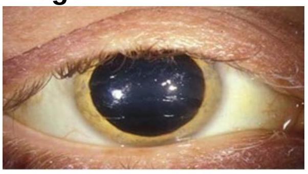
Choose the best method of diagnosis for the clinical sign represented in the image.

What is the first-line treatment for idiopathic thrombocytopenia purpura (ITP)?
Which of the following statements is false regarding beta thalassemia major?
Treatment of chronic phase of CML in pregnant women is -
Multiple episodes of acute chest syndrome are associated with which of the following conditions?
Which of the following is typically not associated with secondary Idiopathic thrombocytopenic purpura?
What is the earliest hematological change following splenectomy?
Which of the following is the MOST characteristic feature of Hemolytic Uremic Syndrome (HUS)?
Explanation: Corticosteroids - The presented symptoms (fever, fatigue, jaundice, anemia with high reticulocytes, elevated indirect bilirubin, and spherocytes) are highly suggestive of warm autoimmune hemolytic anemia (AIHA) [1]. - Corticosteroids are the first-line and often definitive treatment, suppressing the immune system and reducing antibody production against red blood cells. Splenectomy - Splenectomy is considered a second-line treatment option if corticosteroids fail or if the patient requires unacceptably high doses for maintenance. - It removes the primary site of red blood cell destruction but is not the initial definitive treatment. Blood transfusion - Blood transfusion is a supportive measure used to manage severe anemia and stabilize the patient, but it does not address the underlying autoimmune process. - While helping with symptoms, it is not a definitive treatment for the autoimmune destruction of red blood cells. Immunosuppressive therapy - While corticosteroids are a type of immunosuppressive therapy, other forms of immunosuppressive therapy (e.g., rituximab, azathioprine, cyclophosphamide) are typically reserved for cases refractory to corticosteroids and splenectomy [2]. - They are not considered the initial definitive treatment for warm AIHA.
Explanation: ***Multiple myeloma*** - Characterized by **solitary lytic bone lesions** [1], often seen on X-rays, indicating areas of bone destruction [1]. - It is associated with **monoclonal proliferation** of plasma cells [1], leading to both osteolytic lesions and hypercalcemia. *Osteoblast* - Involvement of osteoblasts typically leads to **bone formation**, not lytic lesions. - Conditions like **osteosarcoma** may show lytic lesions, but not from osteoblast activity directly. *Mitral stenosis* - Primarily a **valvular heart disease**, leading to **blood flow obstruction**, not associated with lytic bone lesions. - Symptoms are predominantly related to **cardiac function** rather than bone pathology. *Atherosclerosis* - A vascular condition characterized by **plaque formation** within arteries, affecting blood flow but not leading to bone lesions. - It manifests as cardiovascular diseases, not as **osseous lesions**.
Explanation: ***Serum ceruloplasmin*** - The image shows a **Kayser-Fleischer ring**, a greenish-brown discoloration in the periphery of the cornea, which is pathognomonic for **Wilson's disease**. - **Wilson's disease** is a genetic disorder of copper metabolism characterized by **low serum ceruloplasmin** levels (the primary copper-carrying protein in the blood) and increased copper deposition in various tissues. *Serum copper* - While Wilson's disease involves copper accumulation, **total serum copper** can be normal or even elevated due to widespread tissue damage releasing copper into the circulation, making it an unreliable diagnostic marker on its own. - A low serum copper level can be seen, but it is not as specific as low ceruloplasmin, as much of the copper in serum is bound to ceruloplasmin. *Karyotyping* - **Karyotyping** is used to analyze the number and structure of chromosomes and is primarily indicated for diagnosing chromosomal abnormalities, such as Down syndrome or Turner syndrome. - It is not relevant for diagnosing metabolic disorders like Wilson's disease, which is caused by a mutation in a single gene (ATP7B), not a chromosomal aberration. *PCR* - **PCR (Polymerase Chain Reaction)** is a technique used to amplify DNA sequences and can be used for genetic testing to identify specific mutations. - While genetic testing for the **ATP7B gene** mutation is a confirmatory test for Wilson's disease, it is not the primary or best method for initial diagnosis, especially when classic clinical signs and biochemical markers (like low ceruloplasmin) are present.
Explanation: ***Corticosteroids*** - **Corticosteroids**, such as prednisone or dexamethasone, are the **first-line treatment** for ITP due to their ability to quickly reduce antibody production and improve platelet count by immunosuppression. - They work by **decreasing platelet destruction** in the spleen and improving the integrity of blood vessels. *Blood transfusion* - **Blood transfusions** are not a primary treatment for ITP as they do not address the underlying autoimmune destruction of platelets and donated platelets are rapidly destroyed. - They are reserved for **life-threatening hemorrhage** as a temporary measure to increase platelet count in severe bleeding episodes. *Intravenous immunoglobulins (IVIG)* - **IVIG** is typically used in ITP patients who have **severe bleeding**, are unresponsive to corticosteroids, or require a rapid increase in platelet count for emergent situations. - It works by **blocking Fc receptors** on macrophages, thereby reducing platelet destruction, but is not the initial first-line therapy. *Splenectomy* - **Splenectomy** is considered a **second-line treatment** for ITP, reserved for patients who are refractory to medical therapy or require long-term remission [1]. - It removes the primary site of platelet destruction and antibody production but carries **surgical risks** and is not the initial approach [1].
Explanation: ***Bone marrow iron is depleted*** - In beta thalassemia major, patients typically experience **iron overload** due to repeated blood transfusions, leading to **depleted iron stores** in the bone marrow. - This finding contrasts with conditions where iron stores are preserved or increased, highlighting the pathology of thalassemia. *Growth and development is impaired* - While children with beta thalassemia major may experience growth delay, this statement is **not universally true** for all patients. - Factors such as treatment and iron overload management can influence growth and development outcomes. *Red cell count <4 x 10'2/L* - Normal red cell counts can occur in some patients, particularly in **transfusion-dependent cases**. - Thus, it is not a definitive characteristic of beta thalassemia major, as levels can vary. *Levels of HbA2 < 3.5%* - In beta thalassemia major, HbA2 levels are often **increased**, usually exceeding 3.5% [1]. - This distinguishes it from beta thalassemia trait, where HbA2 is elevated but not to the same extent as in major [1].
Explanation: ***Interferon therapy*** - **Interferon-alpha** is the preferred treatment for chronic phase **CML** in pregnant women because it is not associated with teratogenic effects. - It works by modulating the **immune system** and inhibiting cell proliferation [1]. *Imatinib* - **Imatinib** is a **tyrosine kinase inhibitor (TKI)** that is highly effective for CML [1], [2]. - However, TKIs are generally **contraindicated in pregnancy** due to potential **teratogenic effects** on the developing fetus. *Leukapheresis* - **Leukapheresis** is a procedure used to reduce the **white blood cell count** significantly and rapidly, often in cases of **hyperleukocytosis**. - It is a supportive measure for managing very high cell counts but not a definitive long-term treatment for CML itself, especially in the chronic phase. *Spleenectomy* - **Spleenectomy** may be considered in CML for specific complications such as massive **splenomegaly**, **splenic infarction**, or severe **thrombocytopenia** secondary to splenic sequestration. - It is not a primary first-line treatment for chronic phase CML, particularly in pregnant women, as it doesn't target the underlying **Philadelphia chromosome abnormality**.
Explanation: ***Sickle Cell Disease*** - **Acute chest syndrome** is a leading cause of morbidity and mortality in patients with **sickle cell disease (SCD)**. - It involves new pulmonary infiltrates with fever, chest pain, or respiratory symptoms, often triggered by fat embolism, infection, or vaso-occlusion. *SLE* - While **Systemic Lupus Erythematosus (SLE)** can affect the lungs (e.g., **lupus pneumonitis**, **pulmonary hemorrhage**), it does not typically manifest as recurrent "acute chest syndrome" in the same distinct pattern as SCD [1]. - Lung involvement in SLE is usually due to inflammation or vasculitis, not vaso-occlusive crises [1]. *Sjogren's syndrome* - **Sjogren's syndrome** is primarily an autoimmune disease affecting exocrine glands, leading to dry eyes and mouth. - Pulmonary involvement in Sjogren's, such as **interstitial lung disease** or **bronchiolitis**, is less common and differs from acute chest syndrome. *Bronchiectasis* - **Bronchiectasis** is characterized by permanent dilation and damage to the airways, leading to chronic cough and recurrent infections. - While it involves recurrent lung problems, its pathology and clinical presentation are distinct from the acute, crisis-driven nature of acute chest syndrome.
Explanation: ***Rheumatoid arthritis*** - While rheumatoid arthritis is an **autoimmune disease**, its association with secondary ITP is statistically **less common** compared to other systemic autoimmune conditions or infections. - ITP is primarily associated with conditions that directly lead to the production of **anti-platelet antibodies** or impair platelet production/survival more significantly. *Systemic lupus erythematosus* - **SLE** is a well-established cause of **secondary ITP** due to its widespread autoimmune activity, including the production of autoantibodies against platelets [1]. - Thrombocytopenia is a common hematologic manifestation of lupus. *Hepatitis C infection* - **Hepatitis C virus (HCV) infection** is strongly associated with secondary ITP, often through immune complex formation and direct effects on platelet survival and production. - HCV can induce the production of **anti-platelet antibodies**. *HIV infection* - **HIV infection** is a frequent cause of secondary ITP, with thrombocytopenia often being one of the earliest hematologic abnormalities. - Mechanisms include **direct viral effects on megakaryocytes**, immune-mediated destruction of platelets, and increased platelet consumption.
Explanation: Detailed explanation of hematological changes after splenectomy: ***Leukocytosis and thrombocytosis*** - Following splenectomy, there is an increase in **white blood cells (leukocytosis)** and **platelets (thrombocytosis)**, which is the earliest hematological change observed. - This is due to the loss of splenic function, which normally clears excess blood cells from circulation. *Poikilocytosis* - Poikilocytosis refers to the presence of abnormally shaped red blood cells, which typically indicates an underlying **hemolytic anemia** or **nutritional deficiency** [1]. - This change is **not specific** to splenectomy and may take longer to occur post-surgery. *Presence of Heinz bodies* - Heinz bodies indicate the presence of **denatured hemoglobin** often found in conditions like **G6PD deficiency** or **oxidative stress**. - They are **not a direct consequence** of splenectomy and do not appear as an early hematological change. *Evidence of Howell Jolly bodies* - Howell Jolly bodies are remnants of nuclear material in red blood cells, typically seen after splenectomy due to the spleen's role in **removing such bodies**. - However, their appearance is **later on** compared to leukocytosis and thrombocytosis following surgery.
Explanation: ### a and c - Hemolytic Uremic Syndrome (HUS) is characterized by **microangiopathic hemolytic anemia** along with **thrombocytopenia**, making this correct [1][2]. - It typically results from an **infection**, often associated with **E. coli**, particularly in children [2]. *Positive Coomb's test* - A positive Coomb's test suggests **autoimmune hemolytic anemia**, not HUS. - HUS generally presents with **negative Coomb's test**, indicating hemolysis mechanism is different from autoimmune causes. *Microangiopathic haemolytic anaemia* - While HUS does involve **microangiopathic hemolytic anemia** [2], this alone does not encompass all its characteristics necessary for diagnosis. - HUS must also include **thrombocytopenia**, which is not mentioned in this option [1]. *Thrombocytopenia* - Thrombocytopenia is indeed a feature of HUS, but presenting it alone lacks the context of the complete syndrome [1]. - HUS must also show **microangiopathic hemolytic anemia** to align with the complete clinical picture [2], which is missing here.
Anemia Evaluation and Management
Practice Questions
Hemoglobinopathies
Practice Questions
Thalassemias
Practice Questions
Platelet Disorders
Practice Questions
Coagulation Disorders
Practice Questions
Thrombotic Disorders
Practice Questions
Leukemias
Practice Questions
Lymphomas
Practice Questions
Multiple Myeloma and Plasma Cell Disorders
Practice Questions
Myeloproliferative Neoplasms
Practice Questions
Transfusion Medicine
Practice Questions
Hematopoietic Stem Cell Transplantation
Practice Questions
Get full access to all questions, explanations, and performance tracking.
Start For Free