
Endocrinology — MCQs

On this page
Hypocalcemia is characterized by all except
A 10 day old male pseudohermaphrodite child with 46 XY karyotype presents with BP of 110/80 mmHg. Most likely enzyme deficiency is:
Most common endocrine complication of intracranial radiotherapy is
Hypophosphatemia is caused by-
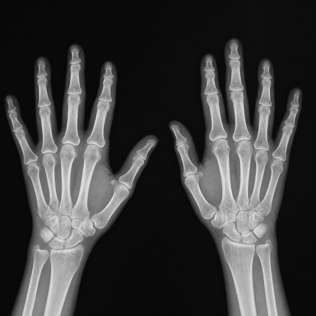
A 65-year-old woman with type 2 diabetes is on hemodialysis for chronic kidney disease. She now presents to the clinic with symptoms of pain in the hands. The symptoms started many months age and are now getting worse. She does not recall any injury to the hands and has not noticed any swelling or redness in the joints.On examination, the joints are normal with no inflammation or tenderness on palpation. There is full range of motion of the fingers and wrists.Lab investigations: calcium (7.2 mg/dL), phosphate (5.5 mg/dL), and PTH level (710 ng/L). (See Figure below) What is the most likely diagnosis?
Gigantism is most commonly caused by:
Mr. Murali has 126 mg/dl of fasting plasma glucose. His venous plasma glucose 2h after ingestion of 75g oral glucose load is 149 mg/dl. This patient comes under which stage of WHO diagnostic criteria of diabetes & intermediate hyperglycemia?
Short 4th metacarpal is a feature of
Excess of calcium intake leads to?
An endocrinologist decided to give a 7 year old boy testosterone therapy and continued it till puberty. This therapy is likely to:
Practice by Chapter
Diabetes Mellitus
Practice Questions
Thyroid Disorders
Practice Questions
Adrenal Gland Disorders
Practice Questions
Pituitary Disorders
Practice Questions
Calcium and Bone Metabolism
Practice Questions
Reproductive Endocrinology
Practice Questions
Lipid Disorders
Practice Questions
Endocrine Hypertension
Practice Questions
Multiple Endocrine Neoplasia
Practice Questions
Obesity and Metabolic Syndrome
Practice Questions
Neuroendocrine Tumors
Practice Questions
Endocrine Emergencies
Practice Questions
Want unlimited practice?
Get full access to all questions, explanations, and performance tracking.
Scan to download app