
Endocrinology — MCQs

On this page
Which of the following drugs is NOT used in the management of SIADH?
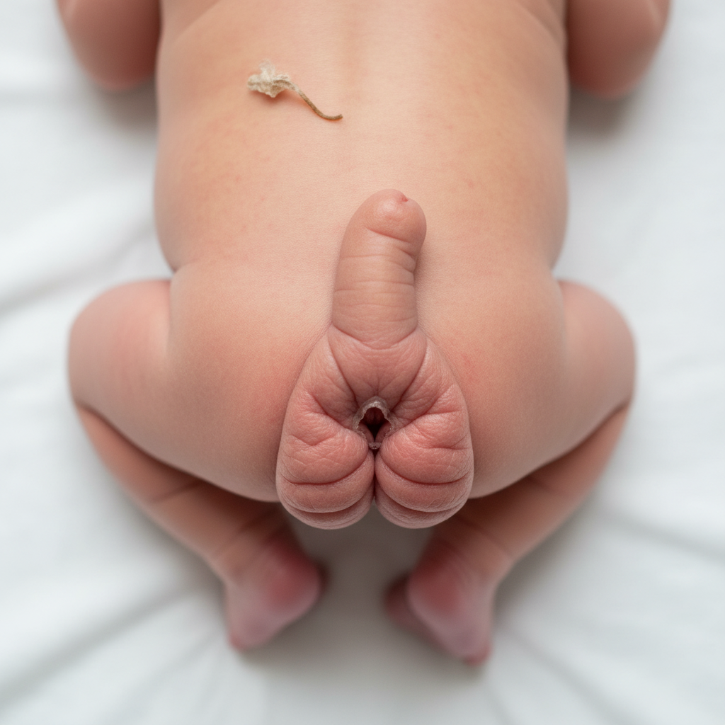
What is the most likely diagnosis in a full-term neonate with electrolyte abnormalities and specific exam findings?
A 72-year-old man is prescribed hydrochlorothiazide for hypertension. Which of the following is the most likely symptomatic side effect?
A patient presents to a clinic with complaints of headache and fatigue. Lab data show serum sodium, 122 mEq/L; serum osmolality, 240 mOsm/L; urine osmolality, 455 mOsm/L. Which condition best correlates with these data?
A 75-year-old woman presents with mild congestive heart failure 6 weeks after a myocardial infarction. She has a history of neck surgery for parathyroid adenoma 5 years ago and her ECG shows slow atrial fibrillation. Her serum calcium is 13.0 mg/dL and urinary calcium is 300 mg/24 h. Examination reveals a small mass in the paratracheal region behind the right clavicle. What is the most appropriate management at this time?
A woman presents with amenorrhea, headache, blurred vision, and galactorrhea. What is the most appropriate investigation?
What is the fasting blood sugar level diagnostic of overt diabetes mellitus?
A 63-year-old male presented with chronic watery diarrhea associated with flushing, characterized by sudden onset deep red or violaceous erythema of the upper body, often with pruritus, lacrimation, and facial edema. The flushing was noticed to be precipitated by exercise, alcohol, stress, and cheese intake. He has a history of wheezing and clinical examination revealed pellagra-like skin lesions. Which of the following is NOT a useful step in the management of this clinical condition?
Which one of the following oral hypoglycemic agents is not an insulin secretagogue?
Reduction in the flow of saliva is not generally seen in which of the following conditions?
Practice by Chapter
Diabetes Mellitus
Practice Questions
Thyroid Disorders
Practice Questions
Adrenal Gland Disorders
Practice Questions
Pituitary Disorders
Practice Questions
Calcium and Bone Metabolism
Practice Questions
Reproductive Endocrinology
Practice Questions
Lipid Disorders
Practice Questions
Endocrine Hypertension
Practice Questions
Multiple Endocrine Neoplasia
Practice Questions
Obesity and Metabolic Syndrome
Practice Questions
Neuroendocrine Tumors
Practice Questions
Endocrine Emergencies
Practice Questions
Want unlimited practice?
Get full access to all questions, explanations, and performance tracking.
Scan to download app