
Endocrinology — MCQs

On this page
What is the commonest thyroid tumor in MEN (Multiple Endocrine Neoplasia)?
Atrial fibrillation is common in which of the following conditions?
Which of the following is true about MEN-I?
A 28-year-old lady has gained 10 kg over 3 years and has oligomenorrhea followed by amenorrhea for 8 months. Her blood pressure is 160/100 mm Hg. Which of the following is the most appropriate investigation?
A young hypertensive patient has serum K+ 2.8 meq/L and elevated plasma aldosterone level with suppressed plasma renin activity. What is the likely cause?
What is the leading cause of adrenal insufficiency?
A 52-year-old woman, diagnosed with osteoporosis via dual-energy x-ray absorptiometry of the hip, presents for management of bone loss prevention. She experienced menopause at age 50 and did not initiate hormone replacement therapy due to a family history of breast cancer. She is concerned about future hip fracture. Which of the following pharmaceutical agents is most appropriate for this patient?
Which of the following statements is true regarding thyroglobulin?
Hyperpigmentation is seen with which of the following hormones?
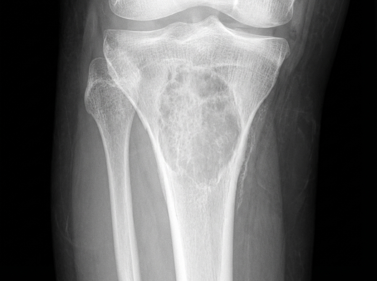
A 40-year-old female presented with severe pain in the left forearm and left ankle following trauma. She reported difficulty walking and swelling in her right upper leg. She also experienced ankle pain and painful ankle joint movements for approximately two years. Relevant abnormal biochemical parameters included: raised serum calcium (11.9 mg/dl), raised alkaline phosphatase (717 U/L), low inorganic phosphorus (1.3 mg/dl), and raised serum parathyroid hormone (1265 pg/l). Protein electrophoresis for M band was negative. An X-ray of the right leg is provided. What is the most likely diagnosis given the clinical presentation and biochemical findings?
Practice by Chapter
Diabetes Mellitus
Practice Questions
Thyroid Disorders
Practice Questions
Adrenal Gland Disorders
Practice Questions
Pituitary Disorders
Practice Questions
Calcium and Bone Metabolism
Practice Questions
Reproductive Endocrinology
Practice Questions
Lipid Disorders
Practice Questions
Endocrine Hypertension
Practice Questions
Multiple Endocrine Neoplasia
Practice Questions
Obesity and Metabolic Syndrome
Practice Questions
Neuroendocrine Tumors
Practice Questions
Endocrine Emergencies
Practice Questions
Want unlimited practice?
Get full access to all questions, explanations, and performance tracking.
Scan to download app