
Endocrinology — MCQs

On this page
Which disorder is associated with thyrotoxicosis but not with hyperthyroidism?
Sarcodiosis can be associated with which of the following?
A young lady presents with tremors, diarrhea, and elevated T4. TSH levels were 8.5 mIU/L. Further examination reveals bi-temporal hemianopia. What is the next step in management?
Thyroid storm can be treated by all the following drugs except?
A patient presents with palpitations, headaches, profuse sweating, and hypertension, raising suspicion for pheochromocytoma. Which diagnostic procedure is NOT typically performed in the evaluation of pheochromocytoma?
All of the following are associated with hypergonadotropic hypogonadism in males, EXCEPT?
Which of the following is NOT a feature of hypercalcemia?
In Sipple syndrome (MEN 2A), which of the following is typically absent?
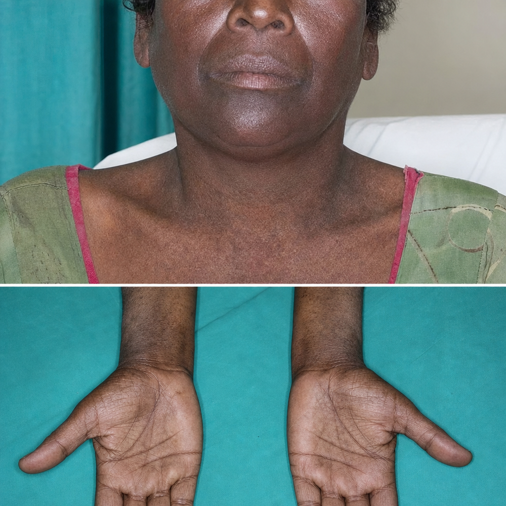
What is the most likely diagnosis in a patient with generalized weakness and the given cutaneous finding?
A patient presents with symptoms of hypoglycemia. Investigations reveal decreased blood glucose and increased insulin levels. A C-peptide assay shows normal levels of C-peptide. What is the most likely diagnosis?
Practice by Chapter
Diabetes Mellitus
Practice Questions
Thyroid Disorders
Practice Questions
Adrenal Gland Disorders
Practice Questions
Pituitary Disorders
Practice Questions
Calcium and Bone Metabolism
Practice Questions
Reproductive Endocrinology
Practice Questions
Lipid Disorders
Practice Questions
Endocrine Hypertension
Practice Questions
Multiple Endocrine Neoplasia
Practice Questions
Obesity and Metabolic Syndrome
Practice Questions
Neuroendocrine Tumors
Practice Questions
Endocrine Emergencies
Practice Questions
Want unlimited practice?
Get full access to all questions, explanations, and performance tracking.
Scan to download app