
Endocrinology — MCQs

On this page
Foot ulcers in diabetes are due to all except which of the following?
What is the most common cause of hypothyroidism in pregnancy?
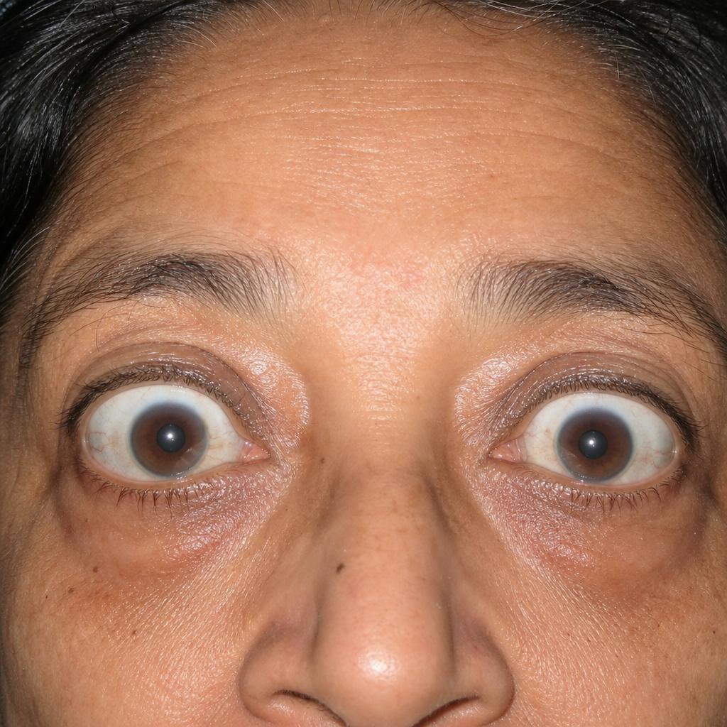
What is the best screening test for pheochromocytoma?
All of the following are seen in MEN 2B except which of the following?
VIPoma is associated with which syndrome?
Hirsutism is seen in all except:
Comment on the diagnosis of this patient with hypokalemic periodic paralysis.
What percentage of cold thyroid nodules are malignant?
A 20-year-old female presents with increasing hair growth on her face and chest, deepening of her voice, and acne over the past year. She has no history of other medical problems. On examination, she has acne, abnormal male pattern balding, and enlargement of her clitoris. Blood tests show normal serum testosterone levels but a markedly elevated level of dihydroepiandrosterone sulphate. What is the most likely diagnosis?
In Klinefelter syndrome, which of the following statements is false?
Practice by Chapter
Diabetes Mellitus
Practice Questions
Thyroid Disorders
Practice Questions
Adrenal Gland Disorders
Practice Questions
Pituitary Disorders
Practice Questions
Calcium and Bone Metabolism
Practice Questions
Reproductive Endocrinology
Practice Questions
Lipid Disorders
Practice Questions
Endocrine Hypertension
Practice Questions
Multiple Endocrine Neoplasia
Practice Questions
Obesity and Metabolic Syndrome
Practice Questions
Neuroendocrine Tumors
Practice Questions
Endocrine Emergencies
Practice Questions
Want unlimited practice?
Get full access to all questions, explanations, and performance tracking.
Scan to download app