Which of the following is NOT a characteristic of tumor lysis syndrome?
What is the earliest finding in diabetic nephropathy?
Conn's syndrome is:
Gynecomastia is seen in which of the following conditions?
Which of the following autoantibodies is NOT commonly found in Graves disease?
A 76-year-old woman presents with a 1-month history of involuntary weight loss, anxiety, and palpitations. She has no prior history of anxiety or palpitations. Her only medical history is hypertension managed with losartan. On examination, her blood pressure is 120/70 mm Hg, heart rate is 100/min and regular. Auscultation reveals normal heart sounds and clear lungs. Physical findings include a thyroid goiter, warm skin, and a fine tremor in her hands. What is the most likely cardiac finding?
A short fourth metacarpal is a characteristic feature of which of the following conditions?
What is the most specific marker for pheochromocytoma?
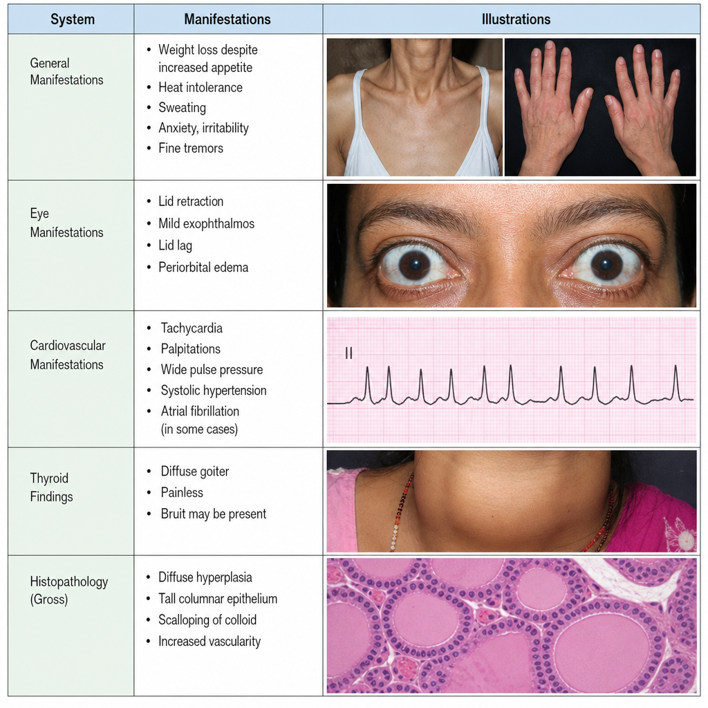
A 35-year-old woman presents with amenorrhea and weight loss despite increased appetite. The history and physical examination reveal exophthalmos, fine resting tremor, tachycardia, and warm, moist skin. What laboratory test for thyroid function would be expected to yield a decreased value?
Galactorrhoea due to ectopic prolactin is seen in which of the following conditions?
Explanation: Enriched Explanation: Tumor Lysis Syndrome (TLS) is an oncologic emergency caused by the massive, rapid breakdown of malignant cells (most commonly in high-grade lymphomas and leukemias) following chemotherapy. This release of intracellular contents into the bloodstream leads to specific metabolic derangements. **Why Hypercalcemia is the Correct Answer:** TLS is characterized by **Hypocalcemia**, not hypercalcemia [1]. As intracellular phosphorus is released, it binds to serum calcium to form calcium phosphate crystals. This "precipitation" leads to a rapid decline in ionized calcium levels, which can cause tetany or seizures. **Analysis of Incorrect Options:** * **Hyperkalemia (A):** Potassium is the primary intracellular cation. Rapid cell lysis floods the extracellular space with potassium, posing a risk for lethal cardiac arrhythmias. This is often the most immediate life-threatening complication. * **Hyperuricemia (C):** The breakdown of nucleic acids releases purines, which are metabolized by xanthine oxidase into uric acid. This can lead to acute uric acid nephropathy and renal failure. * **Hyperphosphatemia (D):** Malignant cells contain significantly higher concentrations of phosphorus than normal cells. Their destruction leads to a surge in serum phosphate, which subsequently drives the secondary hypocalcemia mentioned above. **High-Yield Clinical Pearls for NEET-PG:** * **Cairo-Bishop Criteria:** Used for the diagnosis of laboratory and clinical TLS. * **Prophylaxis/Treatment:** Aggressive hydration is the mainstay. **Allopurinol** (prevents new uric acid formation) or **Rasburicase** (recombinant urate oxidase that breaks down existing uric acid) are used to manage hyperuricemia. * **ECG Changes:** Watch for peaked T-waves (hyperkalemia) and prolonged QT intervals (hypocalcemia).
Explanation: **Explanation:** Diabetic Nephropathy (DN) is a progressive microvascular complication of diabetes. The earliest **clinically detectable** sign of DN is an increase in urinary albumin excretion [1]. **1. Why Option D is Correct:** The natural history of DN begins with **Hyperfiltration** (increased GFR), followed by **Microalbuminuria** (30–300 mg/24h) [1], [2]. However, in the context of standard clinical testing and the options provided, the progression to **Macroalbuminuria** (Urine albumin >300 mg/24 hours), also known as "Overt Nephropathy," marks the definitive clinical stage of the disease. It is the first sign that signifies established glomerular damage and a high risk of progression to end-stage renal disease (ESRD). **2. Why Other Options are Incorrect:** * **A. Shrunken kidney:** Unlike most chronic kidney diseases, diabetic nephropathy characteristically presents with **normal-sized or enlarged kidneys** even in advanced stages. * **B. Fibrin caps:** These are hyaline deposits in the glomerular capillaries. While characteristic of DN, they are **histopathological** findings seen on biopsy, not the earliest clinical finding. * **C. Elevated serum creatinine:** This is a **late finding**. Creatinine typically remains within normal limits until the GFR has dropped by nearly 50% [3]. **High-Yield Clinical Pearls for NEET-PG:** * **Earliest Functional Change:** Hyperfiltration (Increased GFR). * **Earliest Pathological Change:** Thickening of the Glomerular Basement Membrane (GBM) [2]. * **Most Specific Pathological Finding:** Kimmelstiel-Wilson (KW) nodules (Nodular glomerulosclerosis) [2]. * **Screening:** Annual screening for microalbuminuria is recommended starting 5 years after diagnosis in Type 1 DM and at the time of diagnosis in Type 2 DM [1]. * **Management:** ACE inhibitors or ARBs are the drugs of choice as they reduce intraglomerular pressure.
Explanation: **Explanation:** **Conn’s Syndrome** refers to **Primary Hyperaldosteronism**, specifically caused by an aldosterone-secreting adrenal adenoma (the most common cause, followed by bilateral adrenal hyperplasia). In this condition, the adrenal cortex autonomously overproduces aldosterone, independent of the Renin-Angiotensin System. 1. **Why Option A is Correct:** In primary hyperaldosteronism, the pathology lies within the adrenal gland itself. Excess aldosterone leads to increased sodium reabsorption and potassium/hydrogen ion excretion in the distal nephron [2]. This results in the classic triad of **Hypertension, Hypokalemia, and Metabolic Alkalosis**. A key diagnostic feature is a **low plasma renin level** due to feedback suppression by high blood pressure and volume expansion [2]. 2. **Why Other Options are Incorrect:** * **Option B (Secondary Hyperaldosteronism):** This occurs due to external stimuli increasing renin levels (e.g., Renal Artery Stenosis, Congestive Heart Failure, or Cirrhosis) [1]. Unlike Conn’s, both **Renin and Aldosterone are elevated**. * **Options C & D (Hypoaldosteronism):** These involve a deficiency of aldosterone (e.g., Addison’s disease or Hyporeninemic hypoaldosteronism), leading to hypotension and hyperkalemia, which is the clinical opposite of Conn’s syndrome. **NEET-PG High-Yield Pearls:** * **Screening Test:** Plasma Aldosterone Concentration (PAC) to Plasma Renin Activity (PRA) ratio. A **PAC:PRA ratio > 20-30** is highly suggestive. * **Confirmatory Test:** Saline infusion test (failure to suppress aldosterone) or Oral Salt Loading test. * **Management:** Surgical excision for unilateral adenoma (Conn's); Medical management with **Spironolactone or Eplerenone** (Aldosterone antagonists) for bilateral hyperplasia. * **Clinical Hint:** Suspect Conn’s in a young hypertensive patient with unexplained hypokalemia or resistant hypertension.
Explanation: **Explanation:** **Klinefelter’s Syndrome (47, XXY)** is the most common genetic cause of male hypogonadism [2] and a classic cause of gynecomastia. The underlying mechanism involves **primary testicular failure**, where fibrotic changes in the seminiferous tubules lead to low testosterone levels [1]. This triggers a compensatory rise in LH (Luteinizing Hormone), which stimulates Leydig cells to increase the aromatization of testosterone into estradiol [3]. The resulting **decreased testosterone-to-estrogen ratio** leads to the development of breast tissue in approximately 50–80% of patients [1]. **Analysis of Incorrect Options:** * **Secondary Syphilis:** Typically presents with a generalized maculopapular rash (including palms and soles), lymphadenopathy, and condyloma lata. It does not involve the endocrine pathways leading to breast tissue proliferation. * **Tuberculoid Leprosy:** Characterized by hypopigmented, anesthetic skin patches and peripheral nerve enlargement. While **Lepromatous leprosy** can cause gynecomastia (due to orchitis and testicular atrophy), the Tuberculoid form generally does not affect the testes. * **HIV:** While certain antiretroviral therapies (ART) or associated lipodystrophy can mimic breast enlargement, HIV itself is not a primary cause of glandular gynecomastia. **High-Yield Clinical Pearls for NEET-PG:** * **Risk of Malignancy:** Patients with Klinefelter’s have a **20–50 times higher risk** of developing male breast cancer compared to the general population [4]. * **Hormonal Profile:** Characterized by **High FSH/LH** and **Low Testosterone** (Hypergonadotropic Hypogonadism) [1]. * **Clinical Triad:** Small firm testes, gynecomastia, and azoospermia (infertility) [1]. * **Drug-induced Gynecomastia (Mnemonic: DISCO):** Digoxin, Isoniazid, Spironolactone, Cimetidine, Estrogens/Ketoconazole [3].
Explanation: **Explanation:** The hallmark of Graves' disease is the presence of autoantibodies directed against the **TSH Receptor (TSHR)** [1], [2], not the TSH hormone itself. 1. **Why Option D is correct:** Antibodies against the **TSH hormone** are not a feature of Graves' disease. In Graves', the pathology involves **Thyroid-Stimulating Immunoglobulins (TSI)** [1] that mimic the action of TSH by binding to and activating the TSH receptor on thyroid follicular cells, leading to hyperthyroidism and glandular hyperplasia [2]. 2. **Why other options are incorrect:** * **Option A (TSI):** This is the most specific marker for Graves' disease [1]. It is a type of TSH-receptor antibody (TRAb) that directly causes the clinical manifestations of thyrotoxicosis. * **Options B & C (Anti-Tg and Anti-TPO):** While these are the classic markers for Hashimoto’s thyroiditis, they are also found in a significant proportion of Graves' disease patients (Anti-TPO in ~80% and Anti-Tg in ~50%). Their presence indicates a generalized autoimmune thyroid process but they are not the primary drivers of hyperthyroidism in Graves'. **Clinical Pearls for NEET-PG:** * **Most Specific Test:** TSI (Thyroid Stimulating Immunoglobulin) is the most specific for diagnosing Graves' disease and predicting neonatal thyrotoxicosis (as IgG crosses the placenta) [4]. * **Most Sensitive Test:** TSH-receptor antibody (TRAb) assays [1]. * **Triad of Graves:** Hyperthyroidism + Diffuse Goiter + Ophthalmopathy (Exophthalmos) [1], [2]. Pretibial myxedema (Dermopathy) is also highly specific [1]. * **Radioiodine Uptake (RAIU):** Characteristically shows **diffuse, increased uptake**, distinguishing it from thyroiditis (low uptake) or toxic multinodular goiter (patchy uptake) [3].
Explanation: **Explanation:** The clinical presentation of weight loss, anxiety, tremors, and a goiter in an elderly patient is highly suggestive of **Hyperthyroidism** (likely Toxic Multinodular Goiter given the age) [1]. **1. Why Option C is Correct:** Hyperthyroidism induces a hyperdynamic state by increasing the expression of beta-adrenergic receptors and affecting cardiac myocytes directly. In elderly patients, "Apathetic Hyperthyroidism" often presents with cardiac manifestations rather than classic sympathetic symptoms [2]. **Atrial Fibrillation (AF)** is the most common arrhythmia associated with thyrotoxicosis, occurring in 10–15% of patients, particularly those over age 60 [1]. It often starts as paroxysmal AF before becoming persistent. **2. Why Incorrect Options are Wrong:** * **A. Prolonged circulation time:** In hyperthyroidism, the circulation time is actually **shortened** due to increased heart rate and stroke volume (hyperdynamic circulation). Prolonged circulation time is a feature of hypothyroidism or congestive heart failure. * **B. Decreased cardiac output:** Hyperthyroidism causes **increased cardiac output** to meet the high metabolic demands of peripheral tissues. * **D. Pericardial effusion:** This is a classic finding in **Hypothyroidism** (Myxedema), where increased capillary permeability and reduced lymphatic drainage lead to fluid accumulation. **NEET-PG High-Yield Pearls:** * **Most common arrhythmia in Thyrotoxicosis:** Sinus tachycardia [1]. * **Most common "significant" arrhythmia in elderly thyrotoxic patients:** Atrial Fibrillation [2]. * **Thyroid Storm Management:** Propranolol is used not just for tachycardia, but to inhibit the peripheral conversion of T4 to T3 [2]. * **Apathetic Hyperthyroidism:** Always suspect this in an elderly patient with new-onset AF or unexplained weight loss, even if they lack "classic" signs like exophthalmos [3].
Explanation: The correct answer is **Pseudohypoparathyroidism (PHP)**, specifically Type 1a, also known as **Albright Hereditary Osteodystrophy (AHO)**. **1. Why Pseudohypoparathyroidism is correct:** PHP is characterized by **end-organ resistance to Parathyroid Hormone (PTH)**. While PTH levels are high, the body cannot respond to them, leading to hypocalcemia and hyperphosphatemia [1]. Patients with the AHO phenotype (caused by a mutation in the *GNAS1* gene) exhibit distinct physical features, most notably **shortening of the fourth and fifth metacarpals and metatarsals** (Archibald’s sign). This occurs due to premature closure of the epiphyses. Other features include short stature, round facies, and subcutaneous calcifications. The inheritance pattern of AHO involves genetic imprinting, where maternal inheritance of the GNAS1 mutation is associated with PTH resistance and biochemical abnormalities [1]. **2. Why the other options are incorrect:** * **Hypothyroidism:** While it can cause delayed bone age and growth retardation in children (cretinism), it does not typically present with isolated shortening of the fourth metacarpal. * **Hyperthyroidism:** This leads to increased bone turnover and potential osteoporosis, but not structural congenital shortening of specific metacarpals. * **Hypoparathyroidism:** This is a deficiency of PTH secretion. While it shares the biochemical profile of low calcium and high phosphate with PHP, it lacks the phenotypic features of AHO (like the short 4th metacarpal). **Clinical Pearls for NEET-PG:** * **Archibald’s Sign:** A positive sign is when a line drawn tangential to the heads of the 4th and 5th metacarpals passes through the head of the 3rd metacarpal (indicating a short 4th metacarpal). * **Differential Diagnosis:** Short 4th metacarpals are also seen in **Turner Syndrome** (45, XO). * **Pseudopseudohypoparathyroidism (PPHP):** Patients have the AHO phenotype (short metacarpals) but **normal** calcium and PTH levels. It occurs due to paternal inheritance of the same *GNAS* mutation [1].
Explanation: Pheochromocytoma is a catecholamine-secreting tumor arising from the chromaffin cells of the adrenal medulla. Understanding the metabolic pathway of catecholamines is key to identifying the correct diagnostic markers. [1] **1. Why Vanillylmandelic acid (VMA) is the correct answer:** VMA is the **end-stage metabolic byproduct** of both epinephrine and norepinephrine. While plasma metanephrines are considered the most *sensitive* screening test, **24-hour urinary VMA** has historically been regarded as a highly **specific** marker (approaching 95%). Because it is the final product of the degradation pathway, its persistent elevation is a strong indicator of catecholamine overproduction, helping to confirm the diagnosis and minimize false positives. **2. Why the other options are incorrect:** * **B. Catecholamines:** While elevated in pheochromocytoma, plasma catecholamines are highly labile. They can rise significantly due to stress, pain, or posture, leading to lower specificity compared to their metabolites. * **C. 5-Hydroxyindoleacetic acid (5-HIAA):** This is the primary metabolite of serotonin. It is the diagnostic marker for **Carcinoid Syndrome**, not pheochromocytoma. * **D. Serotonin:** Serotonin is secreted by neuroendocrine tumors of the gut (Carcinoid tumors). It plays no role in the diagnosis of adrenal medullary tumors. **Clinical Pearls for NEET-PG:** * **Best Initial Screening Test:** Plasma free metanephrines (highest sensitivity). * **Confirmatory Test:** 24-hour urinary metanephrines and VMA. * **Rule of 10s:** 10% are bilateral, 10% are malignant, 10% are pediatric, and 10% are extra-adrenal (Paragangliomas). [1] * **Associated Syndromes:** MEN 2A, MEN 2B, Von Hippel-Lindau (VHL), and NF-1. [1] * **Management Tip:** Always start **Alpha-blockers** (e.g., Phenoxybenzamine) *before* Beta-blockers to prevent a hypertensive crisis.
Explanation: The clinical presentation of weight loss with increased appetite, amenorrhea, exophthalmos, tachycardia, and fine tremors is a classic description of **Hyperthyroidism**, most likely **Graves' Disease** (suggested by exophthalmos) [2]. **Why TSH is the correct answer:** In primary hyperthyroidism, the thyroid gland overproduces thyroid hormones (T3 and T4). According to the **negative feedback mechanism**, high levels of circulating Free T4 and T3 inhibit the anterior pituitary gland from secreting **Thyroid Stimulating Hormone (TSH)** [3]. Consequently, a suppressed (decreased) TSH level is the most sensitive initial marker for diagnosing primary hyperthyroidism [2]. **Analysis of Incorrect Options:** * **Free T4 (A):** In hyperthyroidism, the thyroid gland is overactive, leading to **increased** levels of Free T4 [3]. * **Radioactive Iodine Uptake (B):** In Graves' disease, the gland is hyperfunctioning and will show an **increased** (diffuse) uptake of iodine to synthesize more hormone [1]. * **T3 Resin Uptake (C):** This test indirectly measures the binding capacity of Thyroid Binding Globulin (TBG). In hyperthyroidism, since TBG binding sites are saturated with endogenous T3/T4, the resin uptake value is **increased**. **NEET-PG High-Yield Pearls:** * **Best Initial Test:** Serum TSH is the single most sensitive screening test for both hyper- and hypothyroidism [3]. * **Graves' Disease Triad:** Hyperthyroidism, Exophthalmos (Ophthalmopathy), and Pretibial Myxedema (Dermopathy) [2]. * **Apathetic Hyperthyroidism:** Seen in elderly patients where typical adrenergic symptoms (tachycardia, tremors) may be absent; they may present only with atrial fibrillation or weight loss. * **Subclinical Hyperthyroidism:** Defined as a low TSH with normal Free T4 and T3 levels [4].
Explanation: Explanation: 1. Why Bronchogenic Carcinoma is Correct: Galactorrhea in the context of malignancy is most commonly a paraneoplastic syndrome caused by the ectopic production of Prolactin. Among the options provided, Bronchogenic carcinoma (specifically Small Cell Lung Cancer and occasionally Squamous Cell Carcinoma) is the most well-documented source of ectopic prolactin secretion [2]. These tumors can bypass the normal hypothalamic-pituitary-prolactin axis, leading to hyperprolactinemia and subsequent galactorrhea. 2. Analysis of Incorrect Options: * Malignant Thymoma: While thymomas are associated with various paraneoplastic syndromes (like Myasthenia Gravis or Pure Red Cell Aplasia), they are not a recognized source of ectopic prolactin. * Medullary Carcinoma Thyroid (MTC): MTC is part of MEN 2 syndromes and typically secretes Calcitonin. While MEN 1 is associated with pituitary prolactinomas, MTC itself does not secrete ectopic prolactin. * Hypernephroma (Renal Cell Carcinoma): RCC is famous for producing ectopic hormones like Erythropoietin (EPO), PTHrP, and Renin [1], but it is not a classic source of ectopic prolactin. 3. High-Yield Clinical Pearls for NEET-PG: * Most common cause of Galactorrhea: Pituitary Prolactinoma (not ectopic) [3]. * Ectopic Prolactin Sources: Besides Bronchogenic carcinoma, other rare sources include Renal Cell Carcinoma (rarely) and Gonadoblastomas. * Drug-induced Galactorrhea: Always rule out Dopamine antagonists (Antipsychotics, Metoclopramide) as they are the most common non-neoplastic cause [3]. * Hook Effect: In cases of extremely high prolactin (>100,000 ng/mL), lab assays may show a falsely low result; serial dilution is required for diagnosis.
Diabetes Mellitus
Practice Questions
Thyroid Disorders
Practice Questions
Adrenal Gland Disorders
Practice Questions
Pituitary Disorders
Practice Questions
Calcium and Bone Metabolism
Practice Questions
Reproductive Endocrinology
Practice Questions
Lipid Disorders
Practice Questions
Endocrine Hypertension
Practice Questions
Multiple Endocrine Neoplasia
Practice Questions
Obesity and Metabolic Syndrome
Practice Questions
Neuroendocrine Tumors
Practice Questions
Endocrine Emergencies
Practice Questions
Get full access to all questions, explanations, and performance tracking.
Start For Free